
|
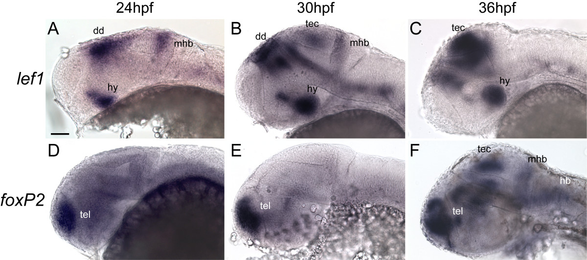
Fig. 1 Sequential expression of foxP2 and lef1 in the CNS during embryogenesis. Whole-mount in situs for lef1 (A-C) and foxP2 (D-F) at 24 hpf, 30 hpf, and 36 hpf. Lateral views, anterior to left, dorsal up; eyes have been removed to facilitate visualization. Scale bar = 50 μm. (Abbreviations: dd, dorsal diencephalon; hb, hindbrain; hy, hypothalamus; mhb, mid-hindbrain boundary; tec, tectum; tel, telencephalon.) (A-C): lef1 is expressed in the hypothalamus, dorsal midbrain, and MHB at 24 hpf, with expression extending to the tectum at 30 hpf. By 36 hpf expression is confined primarily to the hypothalamus and tectum. (D-F): foxP2 is expressed in the tectum and MHB starting at 36 hpf. Earlier expression (24 hpf and 30 hpf) is confined to the telencephalon.