
|
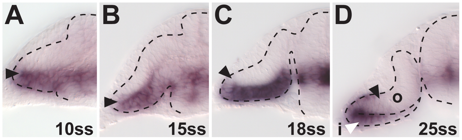
Fig. S6 foxd1 expression in temporal retina progenitors during optic cup formation. (A and B) At 10ss (A) and 15ss (B), foxd1 expression is confined to the ventral optic vesicle leaflet. (C) At 18ss, the first foxd1-positive cells are found at the distal part of the forming outer optic cup layer, indicating the onset of temporal retina progenitor movement into the future neural retina. The whole ventral leaflet is expressing foxd1. (D) At 25ss, foxd1 expression is found in the ventral part of the outer optic cup layer, indicating continued movement. Only the distal part of the inner optic cup layer contains foxd1-expressing cells (white arrowhead), indicating continued movement of temporal progenitors out of this region (black arrowheads: distal/dorsal gene expression limit, dotted lines: neural tube boundary. All images are cross-sections.