
Image
|
Figure Caption
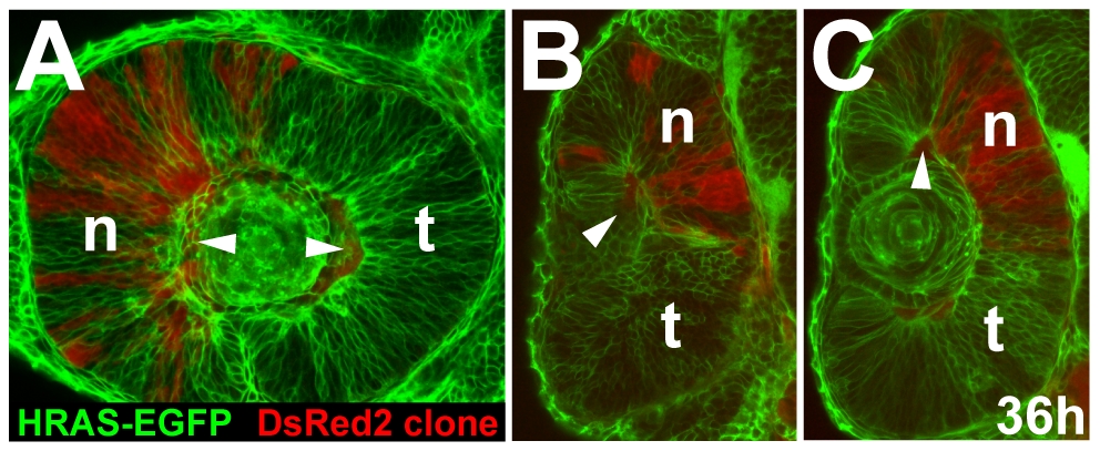
Fig. S5 Nasal restriction of cells from the outer optic cup layer. The DsRed2 cell clone form Figure 3C is restricted to the nasal retina of the HRAS-EGFP host at 36 h. (A) lateral view, (B) optical cross-section at a ventral (B) and medial (C) level along the nasal-temporal axis (arrowheads: autofluorescent blood vessels). n, nasal; t, temporal.
Acknowledgments
This image is the copyrighted work of the attributed author or publisher, and
ZFIN has permission only to display this image to its users.
Additional permissions should be obtained from the applicable author or publisher of the image.
Full text @ PLoS Biol.