
Fig. 6
- ID
- ZDB-IMAGE-091214-22
- Publication
- Picker et al., 2009 - Dynamic coupling of pattern formation and morphogenesis in the developing vertebrate retina
- All Figures
- Figures for Picker et al., 2009
|
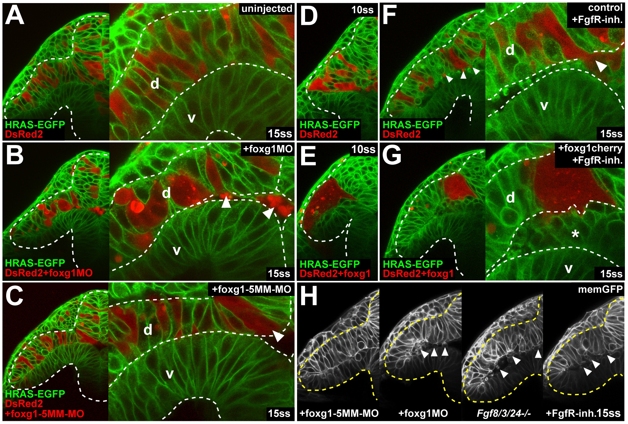
Fig. 6 Foxg1 is required for cell cohesion in the optic vesicle.
(A-C) Live images of a DsRed2-expressing cell clone from a noninjected control donor (A), a foxg1-morpholino-injected donor (B), and a foxg1-mismatch-control-morpholino-injected donor in a Tg(Bactin:HRAS-EGFP)vu119 host embryo (C) at 15ss (right: high magnification view of the dorsal and ventral leaflet in the region of the proximal optic vesicle, arrowheads: delaminating cells). (D and E) Live images of a DsRed2-expressing cell clone from a noninjected control donor (D) and a foxg1-RNA-injected donor (E) in a Tg(Bactin:HRAS-EGFP)vu119 host embryo at 10ss. (F and G) Live images of a DsRed2-expressing cell clone from a noninjected control donor (F) and a foxg1-injected donor (G) in a Tg(Bactin:HRAS-EGFP)vu119 host embryo in the presence of FgfR-inhibitor at 15ss (right: high magnification view of the dorsal and ventral leaflet in the region of the proximal optic vesicle, arrowheads in [F] and asterisk in [G]: delaminating cells). (H) Live images of cell delamination (arrowheads) in a foxg1-mismatch-control-morpholino-injected embryo, a foxg1-morpholino-injected embryo, an fgf8/3/24-/- embryo, and an embryo after FgfR.-inh. treatment at the 15ss stage, coinjected with memGFP RNA (from left to right). Orientation: cross-sections, dorsal to the top and lateral to the left. Dotted lines: neural tube boundary. d, dorsal optic vesicle leaflet; v, ventral optic vesicle leaflet.