
|
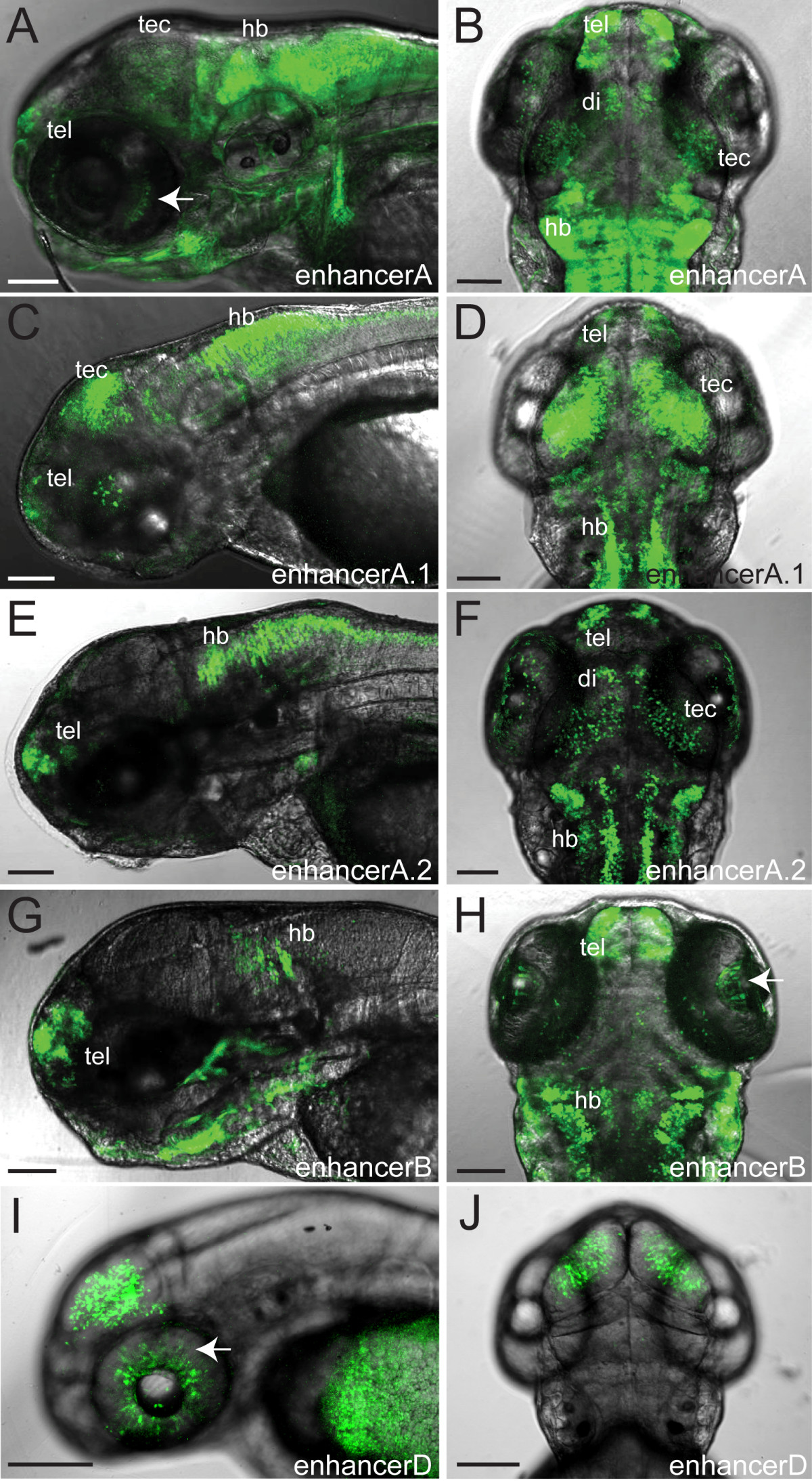
Fig. 5 Confocal live whole-mount images of foxP2 enhancers. Pictures show GFP expression at 72 hpf (except foxP2-enhancerD, taken at 48 hpf). The eye has been removed in panels E and G to facilitate visualization. Scale bar = 100 μm. (A, C, E, G, I): lateral views, anterior left, dorsal up. (B, D, F, H, J): dorsal views, anterior up. (Abbreviations: di, diencephalon; hb, hindbrain; tec, tectum; tel, telencephalon; arrows, GFP-expressing cells in the eye. (A, B) Tg(foxP2-enhancerA:EGFP)zc42; (C, D) Tg(foxP2-enhancerA.1:EGFP)zc44; (E, F) Tg(foxP2-enhancerA.2:EGFP)zc46; (G, H) Tg(foxP-enhancerB:EGFP)zc41; (I, J) Tg(foxP2-enhancerD:EGFP)zc47.