
|
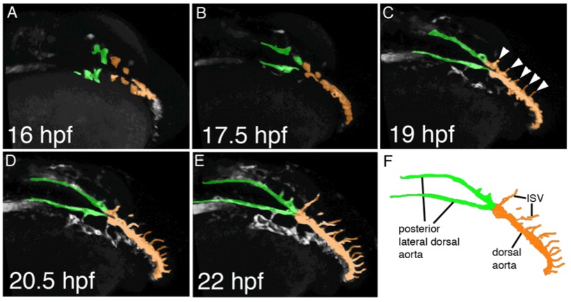
Fig. S2 Still images from 2-photon time lapse movies from wild type Tg(kdrl:egfp) la116 embryos showing endothelial cell movements contributing to lateral dorsal aorta and dorsal aorta formation. A-E, Dorso-lateral view of posterior lateral dorsal aorta formation. Time points are indicated. Endothelial cells that contribute to the lateral dorsal aortae are labeled in green; dorsal aorta is labeled in orange. Note the formation of a Y-shaped structure, consisting of the paired lateral dorsal aortae anteriorly and the single dorsal aorta posteriorly. C, sprouting intersegmental blood vessels are indicated by arrowheads. F, labeled camera lucida image indicating position of vessels in e. ISV – intersegmental blood vessel.