
|
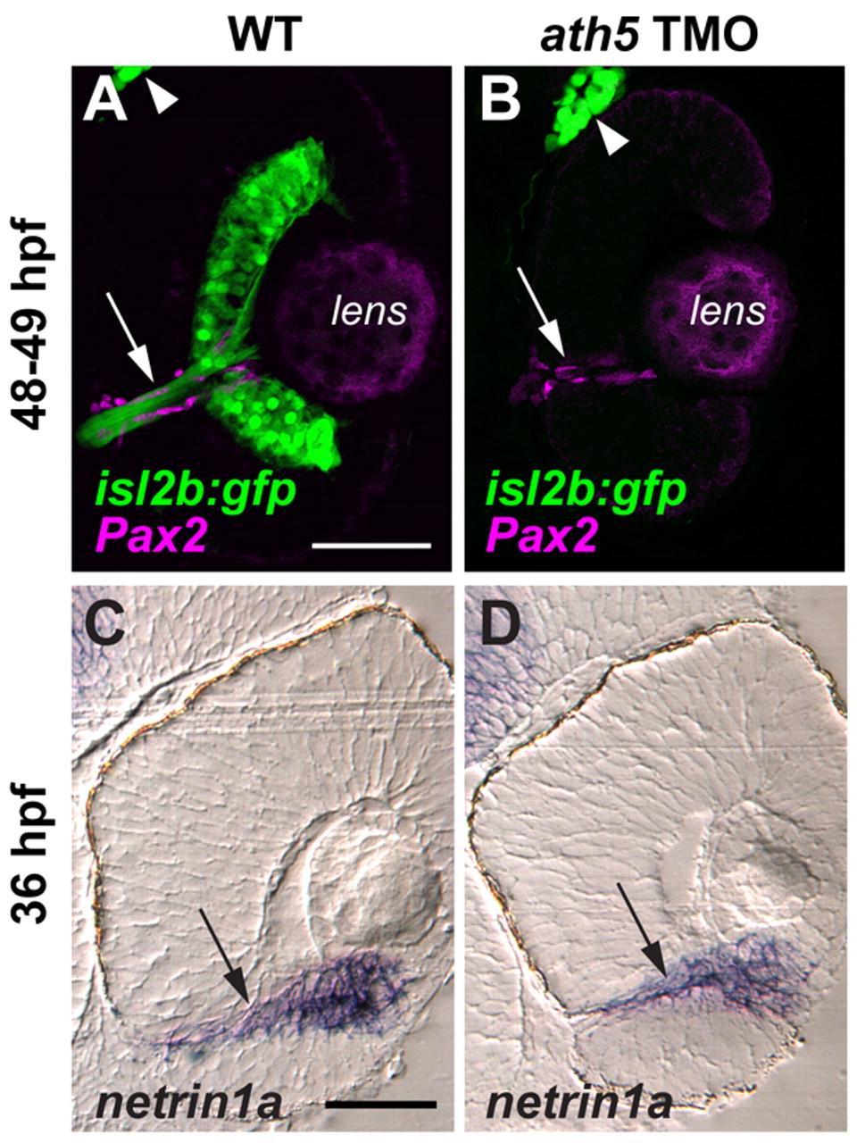
Fig. 3 ath5 MO does not appear to affect the molecular and cellular environment of the optic nerve head or the intraretinal optic nerve. Coronal sections through 48-49 hpf isl2b:GFP (A,B) and 36 hpf non-transgenic (C,D) eyes, from either uninjected embryos (A,C) or embryos injected with high dose ath5 TMO (B,D). (A,B) Pax2a antibody staining (magenta) labels presumptive glial cells which line the intraretinal region of the optic nerve (arrows) in both wild type (A) and ath5 morphants (B). RGCs and their axons (green) are present in wild type (A), but not in ath5 morphants (B). The trigeminal ganglion (arrowheads) is also labeled by isl2b:gfp and serves as a staining control. (C,D) In situ hybridization shows that netrin1a expression (arrows) surrounds the optic nerve and optic nerve head in wild type (C), and is unchanged in ath5 morphants (D). Scale bars: 50 μm.