
|
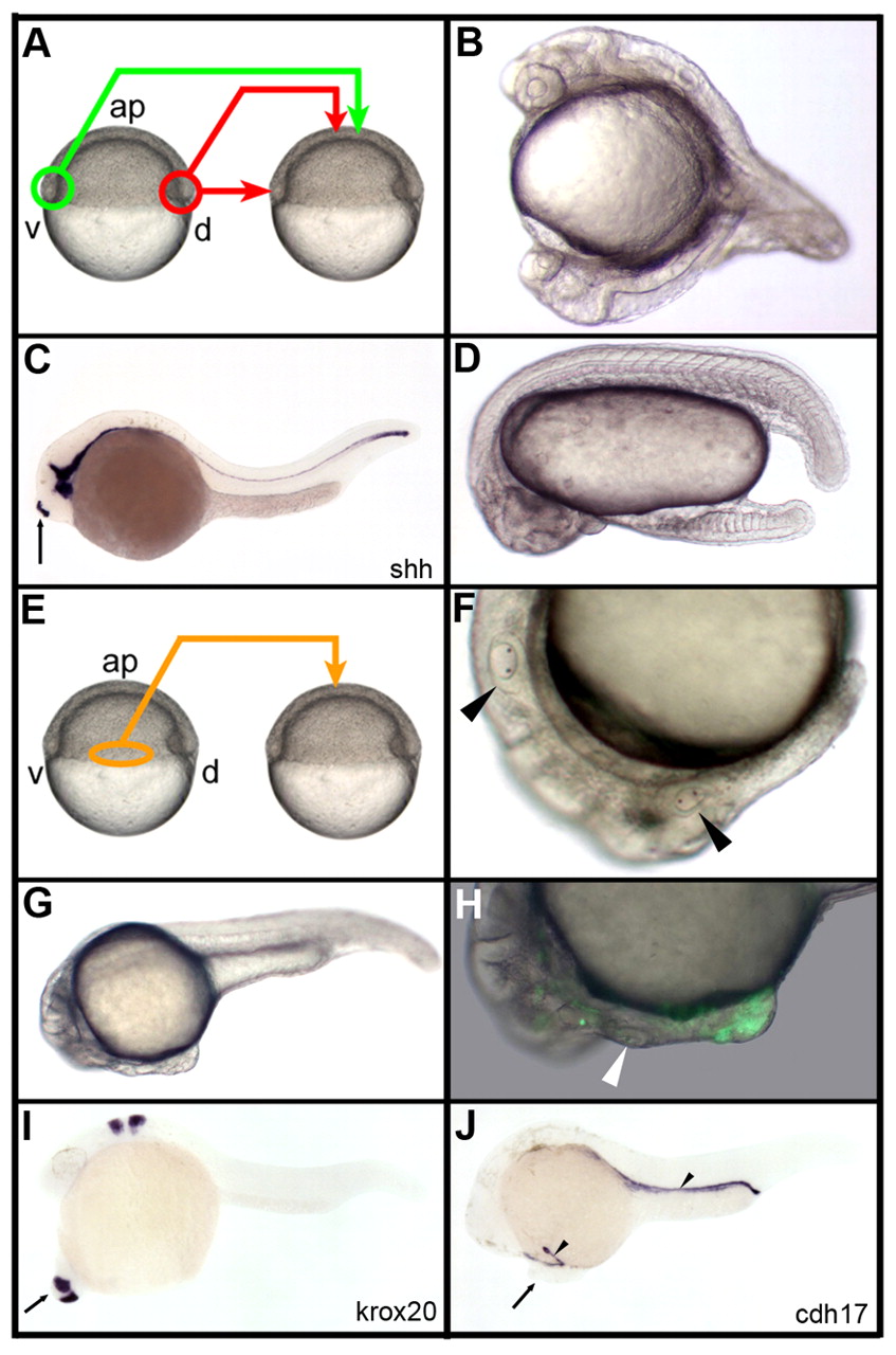
Fig. 1 Organizing properties of the early gastrula margin. (A) Dorsal (red) and ventral (green) marginal grafts of the early gastrula embryo. (B) Transplantation from the dorsal to ventral margin organizes secondary head and trunk. (C) A graft of the dorsal margin at the animal pole induces a short stretch of notochord labeled with the sonic hedgehog shh probe (arrow). (D) A graft of the ventral margin at the animal pole results in the formation of a secondary tail. (E-H) Grafts of the lateral margin (orange, E) organize partial secondary axes containing anterior trunk and posterior head structures (arrowheads in F, otic vesicles in endogenous and secondary axes) that extend from the endogenous head (G) and contain both GFP-labeled cells from the donor, as well as unlabeled cells from the host (H; arrowhead, secondary otic vesicle). (I,J) In situ hybridizations using krox20 and cdh17 RNA probes. Arrow in I,J, secondary axes; arrowhead in J, pronephric ducts; ap, animal pole. B-D,F-J, lateral view, anterior to the left, dorsal to the top; A,E, lateral view, anterior to the top.