
|
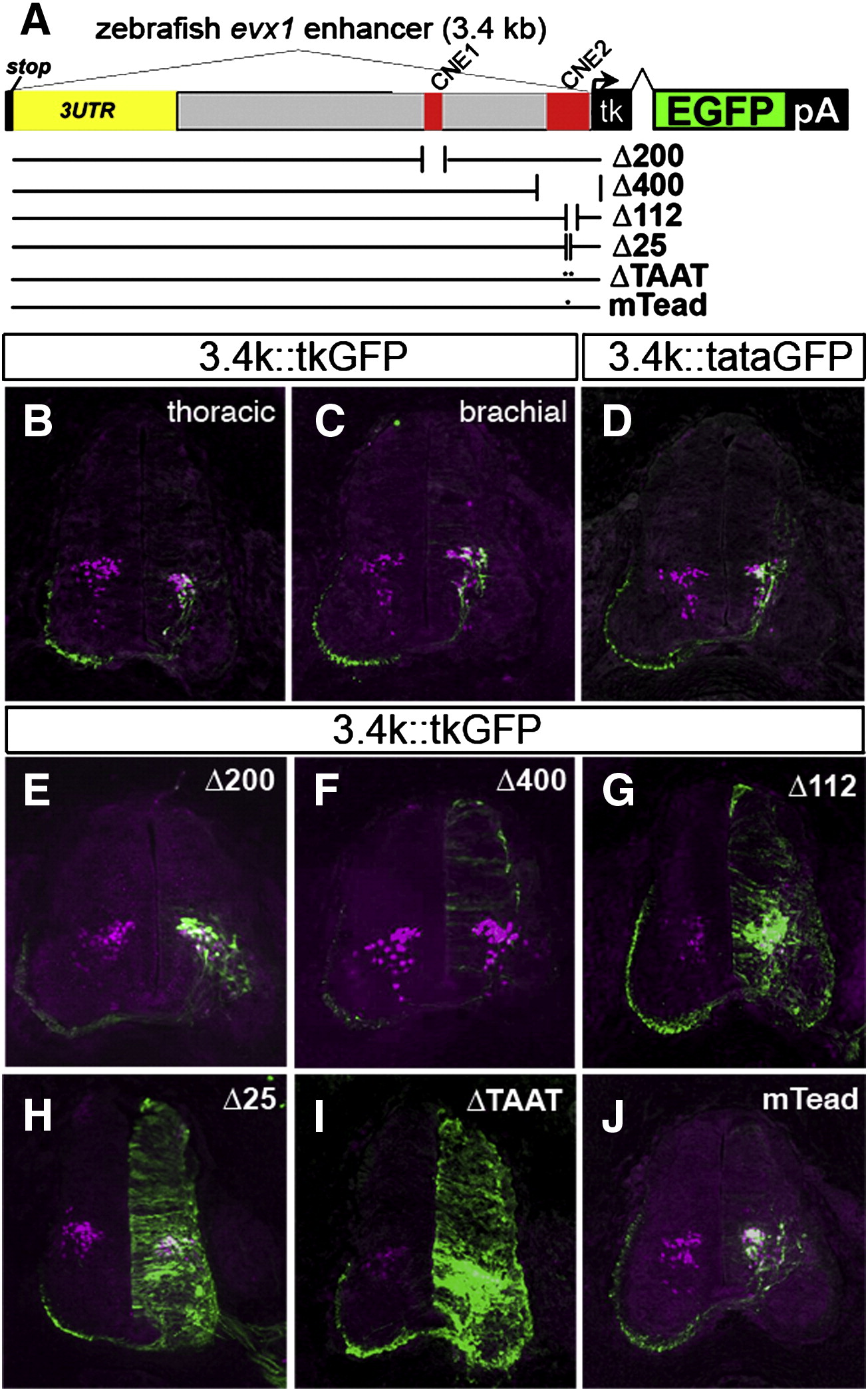
Fig. 5 Deletion analysis of the zebrafish evx1 enhancer in the chick neural tube. (A) Schematic of deletions and mutations in the zebrafish 3.4 kb (3.4 k) downstream fragment driving GFP with a minimal tk promoter in the chick. Deletions Δ200 (NheI/PacI, deletes CNE1), Δ400 (KpnI, deletes CNE2), were generated by digestion and religation. Δ112 (deletes CNE2), Δ25 (deletes 25HCS) and ΔTAAT (deletes HD in 25HCS) were made by PCR deletion mutagenesis (Table S2). In mTead, a putative Tead2 site was mutated (TGAATG to ATTCCG). Sections of electroporated embryos were double labeled with antibodies to GFP and Evx1/Evx2. (B,C) Control sections at thoracic and brachial spinal cord levels. The outline of the spinal cord is drawn as dashed lines. Scale bar: 100 μm. (D) 3.4 kb::tataGFP (with TATA promoter instead) generates an almost identical pattern as with control tk reporter. (E) Δ200 retains V0 labeling and similar specificity to controls. (F) Δ400 eliminates V0-specific labeling but shows low-level background expression. (G) Δ112 retains V0 labeling but shows also widespread expression in the neural tube. (H, I) Δ25 and ΔTAAT also generate widespread GFP expression. (J) Mutation of the putative Tead2 (mTead) site does not alter the pattern of GFP expression. Sections are representative from analysis of 5–9 embryos from each construct.
Reprinted from Developmental Biology, 325(2), Suster, M.L., Kania, A., Liao, M., Asakawa, K., Charron, F., Kawakami, K., and Drapeau, P., A novel conserved evx1 enhancer links spinal interneuron morphology and cis-regulation from fish to mammals, 422-433, Copyright (2009) with permission from Elsevier. Full text @ Dev. Biol.