
Image
|
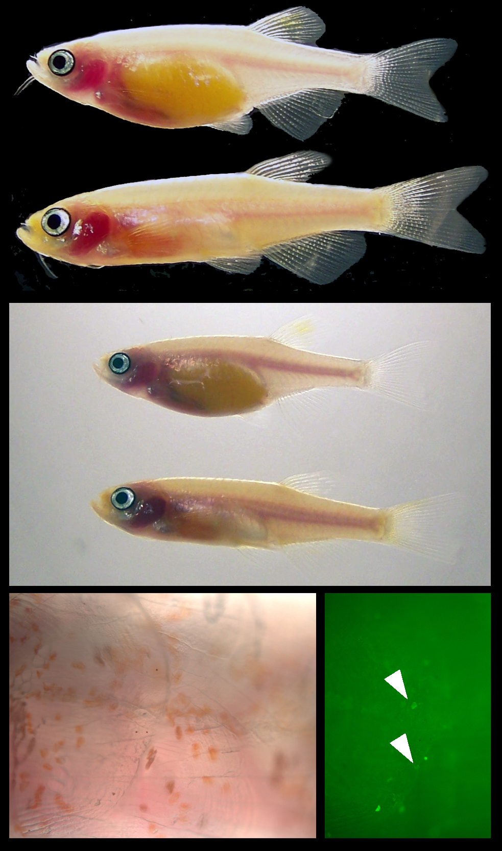
Figure Caption
Fig. 1 Top Panel: Female (top) and male (below) ednrb1 b140/b140;mitfab692/b692 taken with blackfield illumination. Middle Panel: Same male and female ednrb1 b140/b140;mitfab692/b692 - incident and translucent light microscopy combined. Bottom panels: Left: Bright field microscopy, iridophores and melanophores are absent, xantophores (yellow) are present in ednrb1 b140/b140;mitfab692/b692. Right: Fluorescent micrograph with FITC illumination, few iridophores/leucophores (arrowheads, autofluorescent cells) are present in ednrb1 b140/b140;mitfab692/b692 mutants.
Figure Data
Acknowledgments
This image is the copyrighted work of the attributed author or publisher, and
ZFIN has permission only to display this image to its users.
Additional permissions should be obtained from the applicable author or publisher of the image.