
|
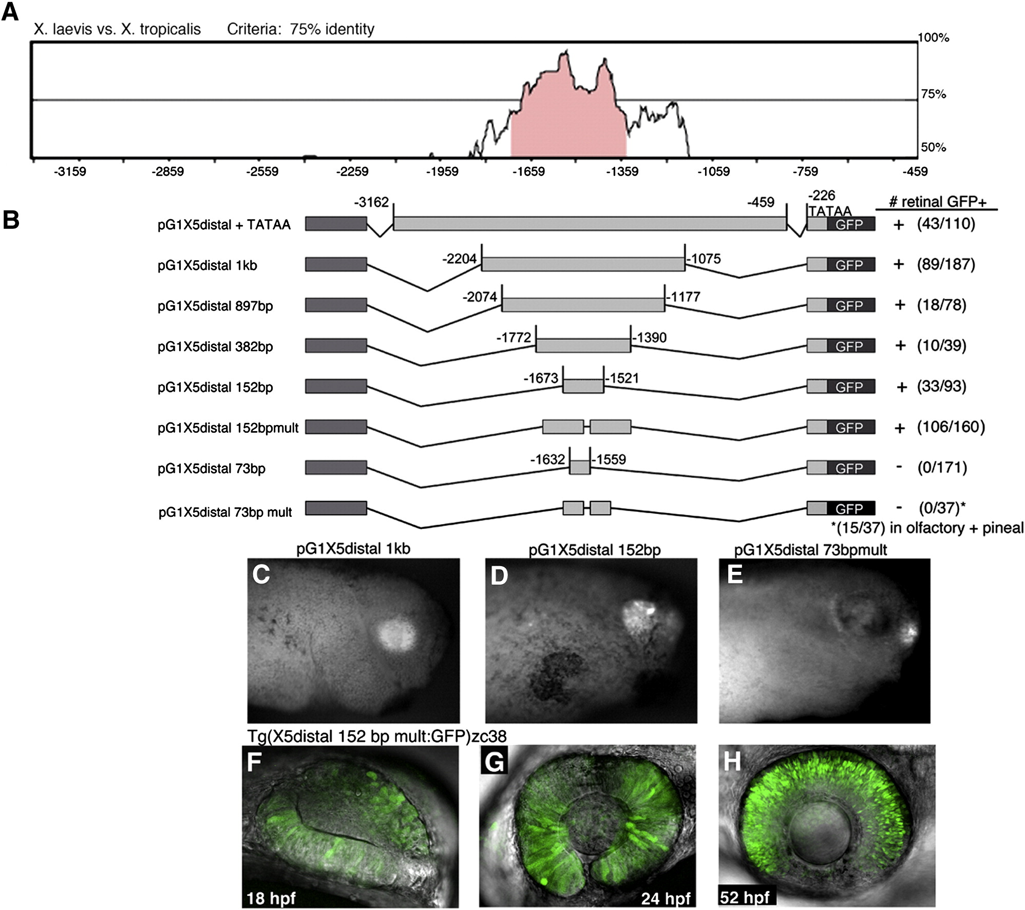
Fig. 1 Identification of a conserved minimal Xath5 distal enhancer that promotes retinal expression in vivo. (A) Pairwise mVISTA (http://genome.lbl.gov/vista/index.shtml) analysis of X. laevis and X. tropicalis Xath5 5′ distal noncoding sequences identifies a 1 kb highly conserved region. (B) A series of nested deletion constructs were generated to identify a minimal 152 bp enhancer that is sufficient to promote retinal specific expression. + strong expression, -/+ very weak expression, - no expression (C) A stage 33 pG1X5 distal 1 kb transgenic embryo shows retinal GFP expression. (D) The 152 bp distal enhancer also drives GFP in the retina. (E) A pG1X5 distal 73 bp transgenic embryo does not express GFP in the retina but only expresses in the pineal gland and olfactory placodes. (F–H) The zc38 transgenic zebrafish line exhibits specific expression in the retina as shown in confocal z-projections at 18 hpf (F), 24 hpf (G), 52 hpf (H) Anterior is to the right in all panels.
Reprinted from Developmental Biology, 326(2), Willardsen, M.I., Suli, A., Pan, Y., Marsh-Armstrong, N., Chien, C.B., El-Hodiri, H., Brown, N.L., Moore, K.B., and Vetter, M.L., Temporal regulation of Ath5 gene expression during eye development, 471-481, Copyright (2009) with permission from Elsevier. Full text @ Dev. Biol.