
|
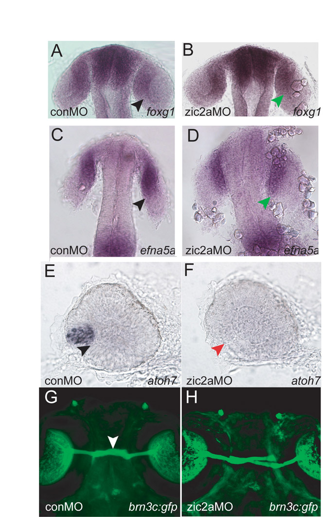
Fig. S7 Gross retinal patterning and retinal neurogenesis appear unaffected in Zic2a knockdown embryos. (A-H) foxg1 expression in the anterior retina in conMO (A, 20/20 embryos) and Zic2a-depleted embryos (B, 21/21 embryos). efna5a expression in the anterior retina in conMO (C, 32/32 embryos, n=2) and zic2a morphants (D, 30/34 embryos, n=2). atoh7 expression marks the ventral retina in conMO-injected embryos (E, 40/46 embryos, n=2). Expression of atoh7 is absent in zic2aMO-injected embryos (F, red arrow, 49/68 embryos, n=2). Tg(pou4f3:gap43-GFP)s356t embryos allow direct visualization of retinal ganglion cells (RGCs) and their axons in living embryos. RGC differentiation and axon guidance (white arrows) are largely normal in control (G) and Zic2a-depleted embryos (H, 7/7 embryos, n=2). A-D show embryos in dorsal view, anterior at the top. E-F show dissected retina at prim-5, anterior to the left. G and H depict live transgenic embryos at 5 dpf imaged by confocal microscopy, ventral view, anterior up.