
|
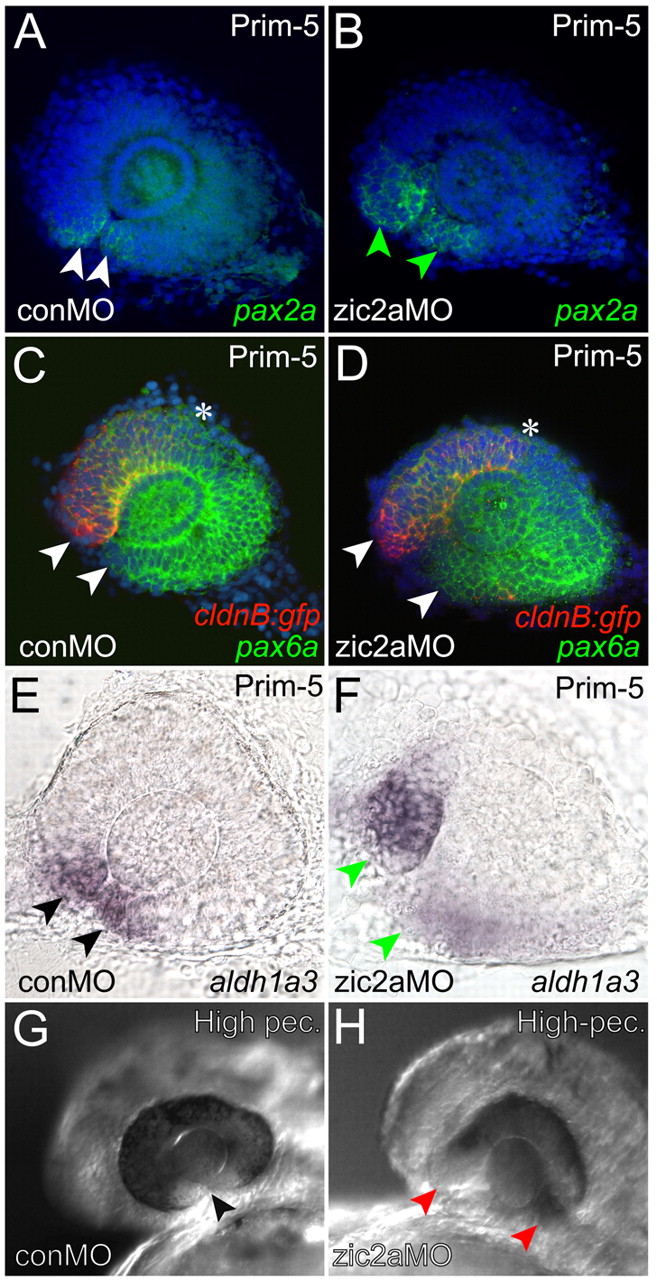
Fig. 6 Ventral retinal defects in zic2a morphants. (A,B) pax2a is normally expressed at the OS-retinal border of control morphants (A) and is expanded into the ventral retina of zic2a morphants (B). (C,D) pax6a expression is seen throughout the retina of Tg(-8.0cldnb:lynGFP)zf106 embryos injected with conMO (C) or zic2aMO (D). Arrowheads in C,D point to the anterior limit of pax6a expression, and asterisks mark the posterior limit of cldnb:gfp expression in the nasal retina. (E,F) aldh1a3 expression is normal in conMO-injected embryos (arrowheads in E, 44/44 embryos, n=2). The zic2a morphant retina fails to close and aldh1a3 expression is expanded (arrowheads in F, 35/38 embryos, n=2). The choroid fissure is closed in uninjected embryos (arrowhead in G), but open in zic2a morphants (red arrowheads in H, 21/28 embryos, n=2). A-F show dissected retinae at prim-5, anterior to the left. A-D are confocal z-stacks. Embryos in G,H are at the high-pec stage