
Image
|
Figure Caption
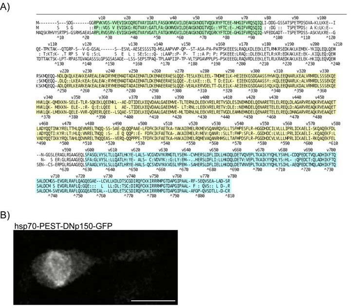
Fig. S1 A) Alignment of Dnct1 from zebrafish (top sequence) versus human (bottom sequence) for the region of the dominant negative protein. Alignments were generated in MegAlign (DNAstar, V6) using the Lipman-Pearson protein algorithm with 0 gap penalty. Overall sequence identity between the proteins is 65%. within specific domains, however, identity is higher: Microtubule binding domain/Cap-Gly region, 86% identity (green); dynein motor binding domain, 84% identity (yellow); dynein intermediate chain binding domain, 80% identity (blue).
B) The dominant negative p150-GFP labels spindle structures at mitosis.
Acknowledgments
This image is the copyrighted work of the attributed author or publisher, and
ZFIN has permission only to display this image to its users.
Additional permissions should be obtained from the applicable author or publisher of the image.
Reprinted from Cell, 138(6), Norden, C., Young, S., Link, B.A., and Harris, W.A., Actomyosin is the main driver of interkinetic nuclear migration in the retina, 1195-1208, Copyright (2009) with permission from Elsevier. Full text @ Cell