
|
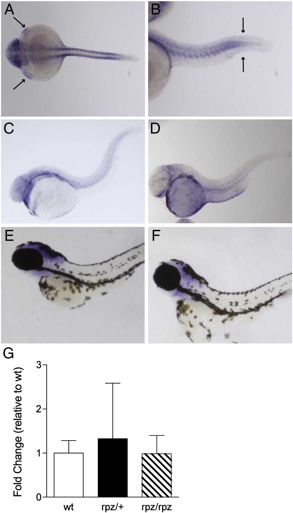
Fig. 4 Expression pattern and qPCR of rpz. (A) Dorsal view of an ISH for rpz in a wild type embryo at 24 hpf. Transcript is clearly seen in the somites, pectoral fin buds (small arrows) and head. (B) In a lateral view, transcript is seen in the somites and in the tail bud adjacent to the fin fold (large arrows). (C–F) Lateral views of wild type embryos at 48 hpf (C), 72 hpf (D), 80 hpf (E), and 120 hpf (F) following ISH for rpz. Expression is beginning to decrease at 48 hpf, and further still at 72 hpf, but the general distribution is unchanged from 24 hpf embryos. By 80 hpf rpz expression is localized exclusively to the head (E) and by 120 hpf expression is almost completely absent (F). (G) Quantitative RT-PCR on wild type, heterozygous and homozygous rapunzel embryos reveals no differences in rpz transcript abundance. Data are normalized to the wild type embryo. Data represent means ± SD.
Reprinted from Developmental Biology, 334(1), Green, J., Taylor, J.J., Hindes, A., Johnson, S.L., and Goldsmith, M.I., A gain of function mutation causing skeletal overgrowth in the rapunzel mutant, 224-234, Copyright (2009) with permission from Elsevier. Full text @ Dev. Biol.