
Fig. 8
- ID
- ZDB-IMAGE-091012-8
- Publication
- Jacoby et al., 2009 - The zebrafish dystrophic mutant softy maintains muscle fibre viability despite basement membrane rupture and muscle detachment
- All Figures
- Figures for Jacoby et al., 2009
|
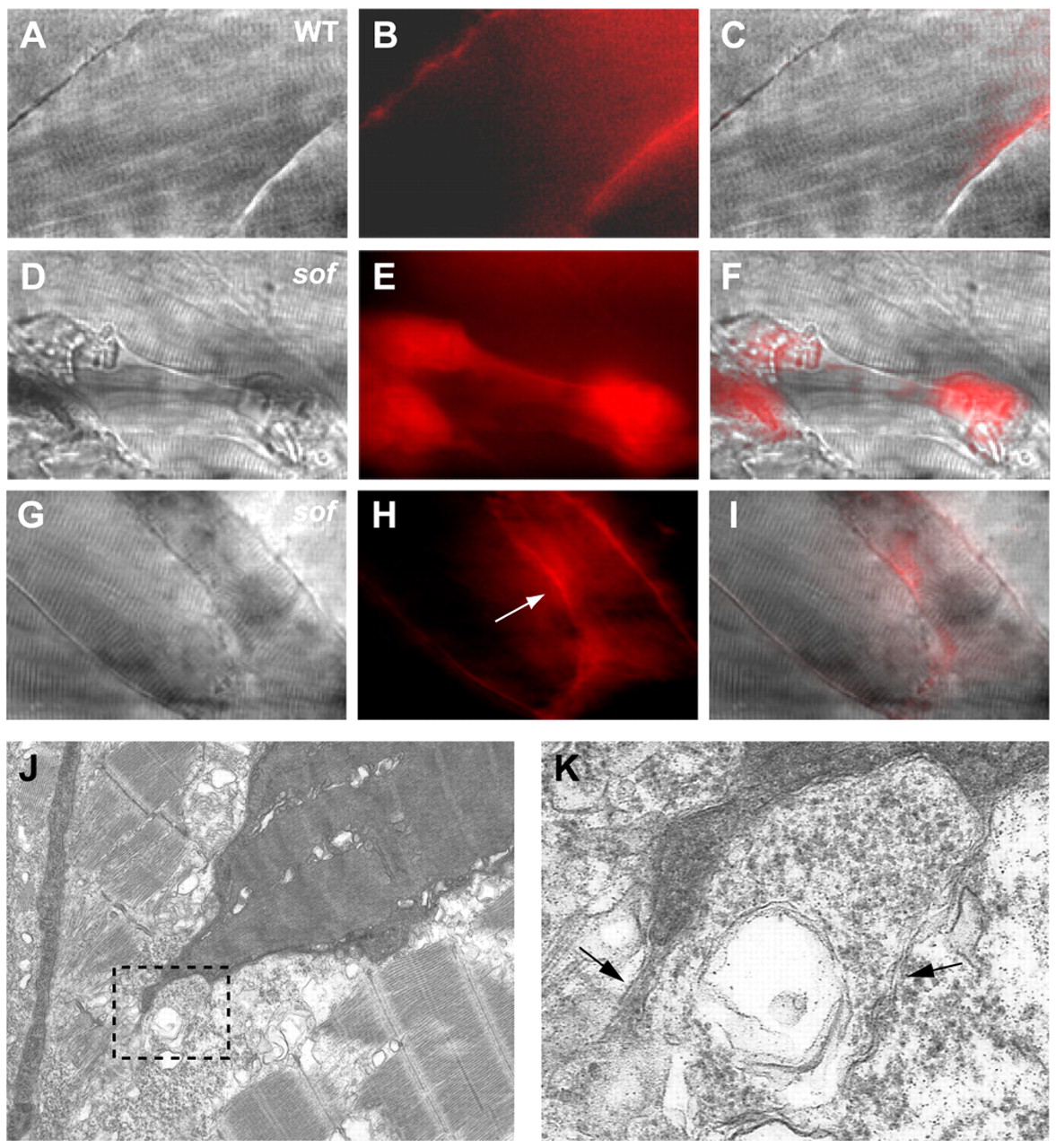
Fig. 8 Sarcolemmal rupture correlates with the mode of fibre detachment in sof muscle. (A-C) Evans Blue is excluded from the myotome of wild-type embryos. (D-F) The dye infiltrates a subset of damaged fibres in sof embryos before complete detachment. The fibre depicted still spans the somite, but the contractile cytoskeleton has collapsed in the centre. (G-I) Evans Blue permeates an EFT (arrow in H) but does not enter the fibres attached to it. (J) Electron micrograph of a retracting fibre in a sof embryo at 72 hpf. The vertical myoseptum is to the left of the fibre. (K) A magnified view of the boxed area in J showing trailing sarcolemma at the newly detached tip of the fibre (arrows). (A,D,G) DIC images, (B,E,H) fluorescence images, (C,F,I) merge of DIC and fluorescence images. All panels show lateral views with anterior to the left.