
|
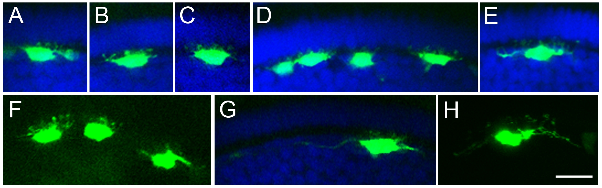
Fig. S2 Morphology of example horizontal cells in Ptf1a:GFP DNA injected embryos (120 hpf). Micrographs showing single images or extended focus views. The nuclear stain DAPI was used to reveal the retinal layers. Horizontal cells have somas in the outer inner nuclear layer immediately adjacent to the outer plexiform layer. Different horizontal cell types have more or less elongated somas and the dendritic trees of different types can extend to form relatively smaller (for example, (A, B, C)) or larger (for example, (E, H)) arbors. Pattern of dendritic tips could not be distinguished in this vertical view and horizontal cells were not further classified, although based on the morphology shown in this vertical view, examples of the previously described types of horizontal cells could be found. Some well-labelled cells also had a distinct axon (G). Scale bar = 20 μm.