
Fig. 5
|
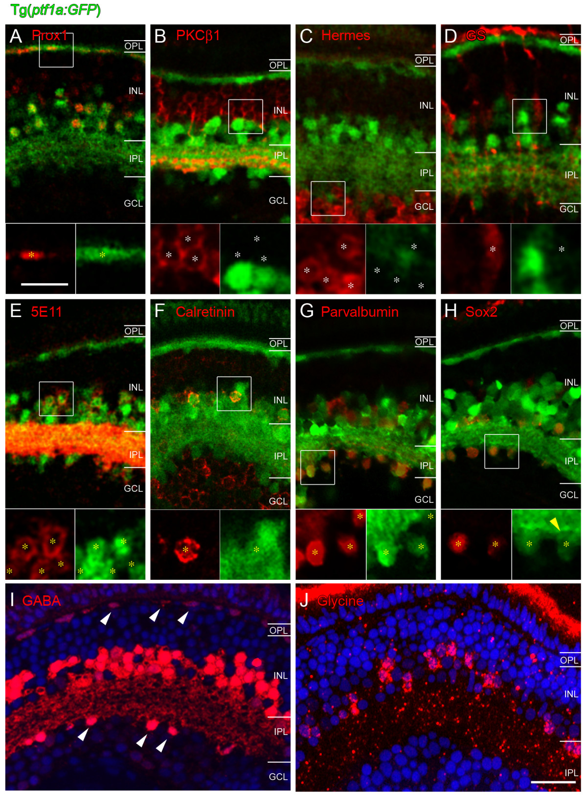
Fig. 5 Immunohistochemical characterization of GFP-expressing cells in Tg(ptf1a:GFP) embryos at 120 hpf. Antibodies used are shown in each panel and appear in the red channel. Insets show higher magnification in which all red (antibody marker) cells are marked by asterisks. White asterisks show red cells that are Ptf1a:GFP negative (B-D), yellow asterisks show red cells that are Ptf1a:GFP positive (A, E-H). (A) All horizontal cells as identified by Prox1 immunoreactivity also express Ptf1a:GFP. Prox1 also weakly labels bipolar cells, which do not express Ptf1a:GFP and a subpopulation of amacrine cells, which does co-label with the Ptf1a:GFP. (B-D) Ptf1a:GFP expressing cells do not colocalise with the bipolar cell marker PKCβ1, the ganglion cell marker Hermes (including the displaced amacrine cells in the ganglion cell layer) nor the Müller cell-specific marker glutamine synthetase. (E-H) In contrast, Ptf1a:GFP-expressing amacrine cells colocalise with the pan-amacrine marker 5E11, and with the amacrine subpopulation markers calretinin, parvalbumin and Sox2. Calretinin (F) is also expressed in ganglion cells and does not label the displaced amacrine populations. (I, J) GABA and glycine staining in plastic sections reveal that some cells in the amacrine layer of the INL express GABA and some express glycine. Some cells in the outermost INL (horizontal cells) and ganglion cell layer (displaced amacrine cells) label with GABA (white arrowheads), but never glycine. The nuclear stain DAPI was used to label the retinal layers. GABA, γ-aminobutyric acid; GCL, ganglion cell layer; GS, glutamine synthetase; INL, inner nuclear layer; IPL, inner plexiform layer; OPL, outer plexiform layer; PKC, protein kinase C. Scale bar in (J) = 20 μm and applies to (A-J); scale bar in the inset of (A) = 10 μm and applies to insets in (A-H).