
|
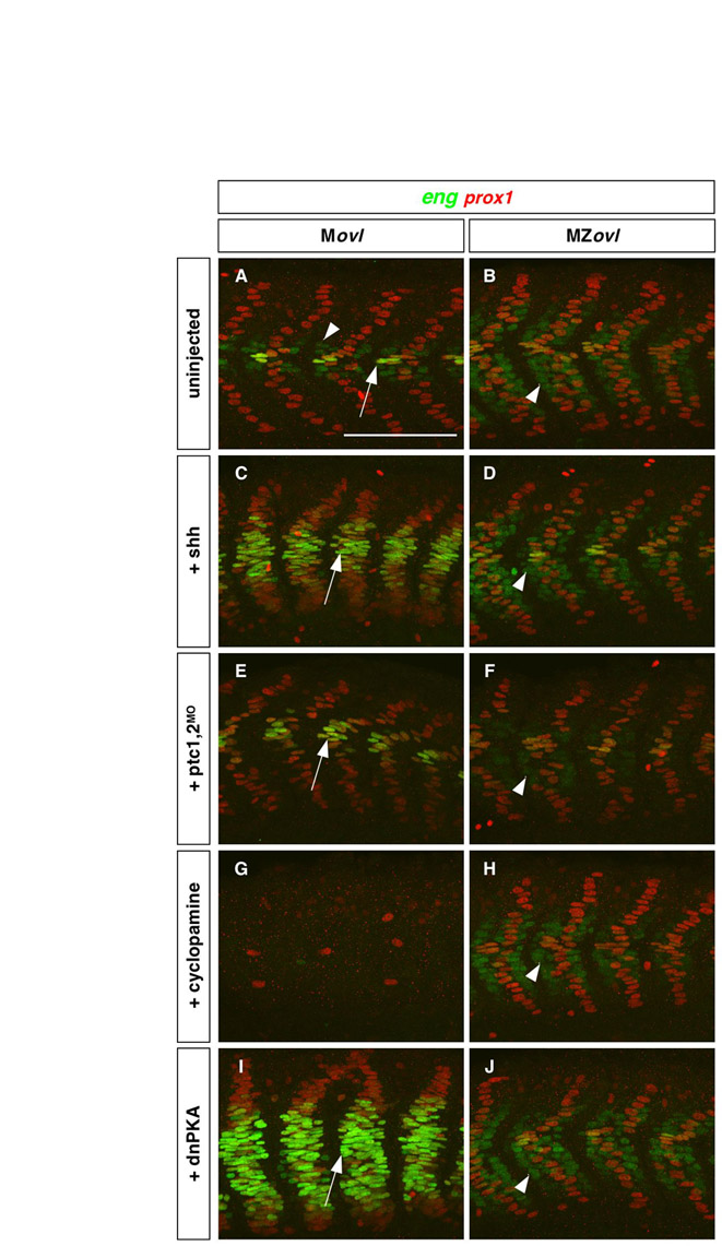
Fig. S6 Epistasis analysis of cilia and Hh signaling in somite patterning. (A-J) Movl control embryos (a,c,e,g,i) and MZovl mutants (b,d,f,h,j) were stained for the expression of eng (green) and prox1 (red) following different manipulations. Ectopic expression of shh mRNA (C), depletion of ptc1,2 using morpholinos (E), and expression of dnPKA mRNA (I) in Movl embryos induce ectopic muscle pioneer cells (prox1+, strong eng+, arrows) and superficial slow fibers (prox1+) as compared with uninjected controls (a), whereas treatment with cyclopamine at 100 μM (g) blocks the formation of most muscle pioneers, superficial slow fibers and medial fast fibers (prox1-, weak eng+, arrowhead in A). By contrast, MZovl embryos are insensitive to these manipulations and are indistinguishable from untreated MZovl embryos, in which medial fast fibers (arrowheads) are expanded (b,d,f,h,j). Lateral views at 24 hpf. Scale bar: 100 μm.