
|
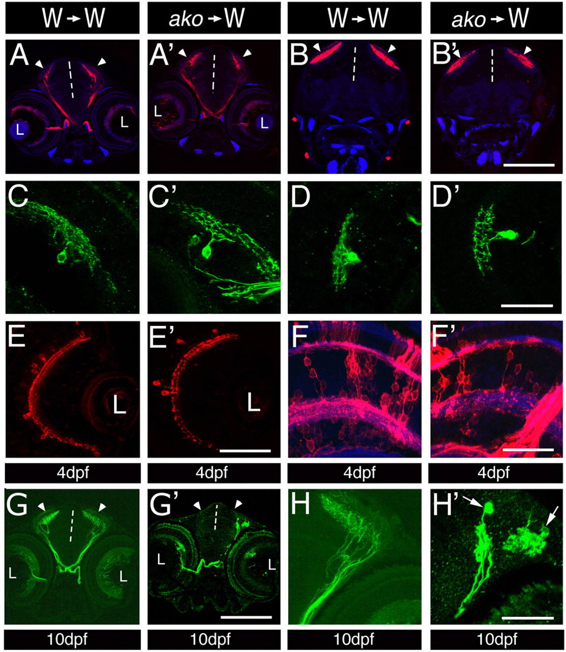
Fig. 4 Cell-autonomous aspects of ako function in neuronal differentiation. (A-H′) Transverse sections through mosaic retinae at 4 (A-F′) and 10 (G-H′) dpf. Donor cells were derived from the following fluorescent protein transgenic lines: Tg(brn3c:mGFP) in A-D′ and G-H′ to visualize ganglion cells; Tg(pax6-DF4:mGFPs220) in E,E′ to isualize amacrine cells; and Tg(pax6-DF4:mCFPq01) in F,F′ to reveal the morphology of bipolar cells. Host tissue is unlabelled. Sections in A,A′ are anterior to those in B,B′. The genotypes of donor and host larvae are indicated above each column. No differences are observed between wild-type and mutant ganglion, bipolar or amacrine cells at 4 dpf. (G,H) Donor-derived wild-type ganglion cells differentiate densely branched processes in the optic tectum of wild-type hosts at 10 dpf. (G′,H′) By contrast, donor-derived akojj50 mutant cells differentiate few axons, which frequently do not branch, or exhibit an irregular branching pattern. In some cases, swellings form near the termini of akojj50 mutant axons (arrows in H′). Vertical dashed lines indicate the midline; arrowheads indicate the optic tecta; L, lens. Scale bars: 150 μm in A-B′,G,G′; 40 μm in C-D′,H,H′; 50 μm in E,E′; 30 μm in F,F′.