
Image
|
Figure Caption
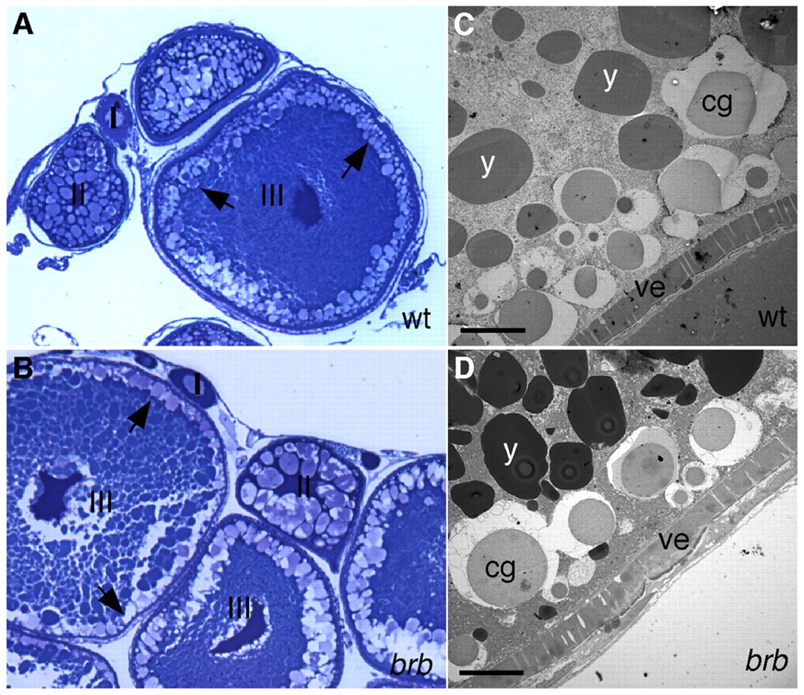
Fig. 4 Oogenesis appears normal in brom bones mutant females. (A,B) Stages I to III of oogenesis appear normal in brom bones mutant females (B, n=3 ovaries). Arrows indicate the location of CGs. (C,D) Transmission electron micrographs of stage IV oocytes. The morphology of cortical granules (cg), yolk globules (y) and vitelline envelope (ve) appear similar in wild-type (C) and mutant (D) oocytes. Scale bar: 10 μm.
Acknowledgments
This image is the copyrighted work of the attributed author or publisher, and
ZFIN has permission only to display this image to its users.
Additional permissions should be obtained from the applicable author or publisher of the image.
Full text @ Development