
|
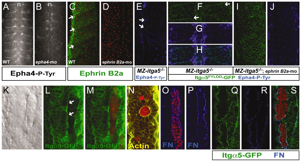
Fig. 5 Eph/Ephrin signaling can induce Itgα5 clustering and FN matrix assembly. (A-D) Phosphorylated Epha4 (Epha4-P-Tyr) localizes to somite boundaries and the notochord (n) in wild-type zebrafish embryos (A) but is substantially reduced in epha4 morpholino-injected embryos (B). Ephrin B2a (green) is expressed in a graded fashion in the posterior somites (arrows) of wild-type embryos (C) but is absent in ephrin B2a morpholino-injected embryos (D). Nuclei are red. (E) Epha4-P-Tyr localizes to transient somite boundaries (arrows) in MZ itga5-/- embryos. (F-H) Epha4-P-Tyr blue) colocalizes with ligand-independent Itgα5FYLDDD-GFP clustering (arrows) in MZ itga5-/- embryos. (I,J) Morpholino knockdown of ephrin B2a abolishes ligand-independent Itgα5FYLDD-GFP clustering. (K-M) epha4-expressing clones (red) in a host with no epha4 but with broadly expressed ephrin B2a. Eph/Ephrin signaling can induce a border (K) and Itgα5-GFP clustering (L,M) around the donor clone. Note that Itgα5-GFP in the host cells is largely localized to the border with the clone (arrows), not along the lateral cell cortices. (N) A rosette with polarized host cells surrounding an epha4-expressing clone. Actin fibrils concentrate along the `basal′ surface of the host cells, similar to polarization along somite boundaries. (O,P) FN matrix is assembled around the epha4-expressing clone. (Q-S)A dnepha4-expressing clone (red) induces Itgα5-GFP clustering and FN matrix assembly. C,D,H,M,N,O,S are overlays.