
|
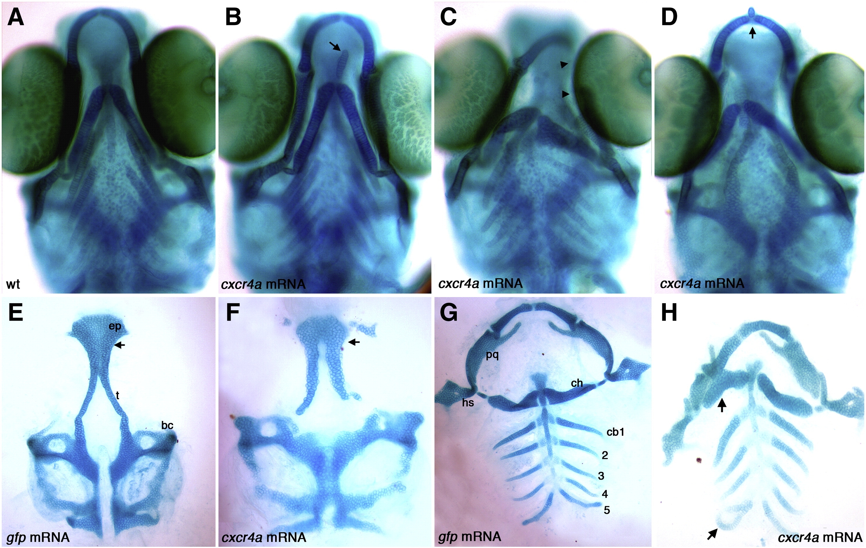
Fig. S4 Overexpression of cxcr4a causes ectopic cartilage formation. Whole-mount (A–D) and dissected (E–H) alcian blue stained cartilage at 6.5 dpf, anterior to the top. Wildtype (A) and gfp mRNA injected controls (E,G) and examples of cxcr4a mRNA injected embryos (B–D,F,H). Injection of 100 pg cxcr4a mRNA results in small ectopic cartilages often located near the Meckel′s and ceratohyal cartilages (B, D; arrow). We also often observe unilateral loss of Meckel′s cartilages and abnormal palatoquadrate elements (C; arrowheads). Injection of 250 pg cxcr4a mRNA results in abnormal hyosymplectic elements (hs), fusions of the Meckel′s and ceratohyal cartilages, as well as fusions of the posterior ceratobranchial elements (H, arrow) as compared to control embryos injected with gfp mRNA (G). Injection of 100 pg of cxcr4a mRNA causes ectopic cartilages and neurocranium defects with a reduction of the trabeculae (t) and a cleft within the ethmoid plate (ep; F; arrow) as compared to control embryos injected with gfp mRNA (E; arrow at same location). bc, basicapsular commissure; cb1–5, ceratobranchial cartilage 1–5; ch, ceratohyal; hs, hyosymplectic; pq, palatoquadrate; ep, ethmoid plate; t, trabeculae.
Reprinted from Developmental Biology, 333(1), Olesnicky Killian, E.C., Birkholz, D.A., and Artinger, K.B., A role for chemokine signaling in neural crest cell migration and craniofacial development, 161-172, Copyright (2009) with permission from Elsevier. Full text @ Dev. Biol.