
Fig. 2
|
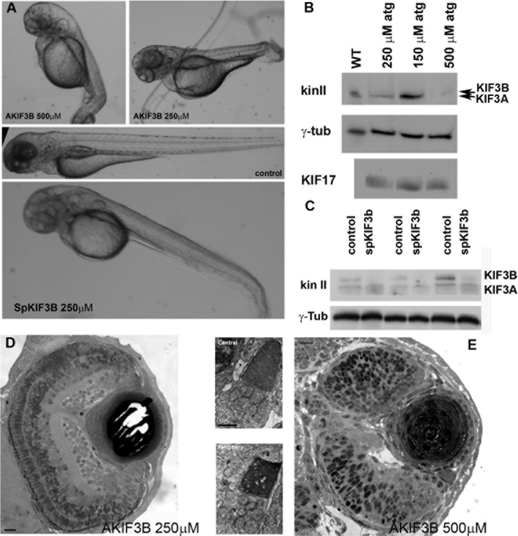
Fig. 2 Disruption of kif3B expression by an antisense morpholino oligonucleotide. A: (Upper panel) Images of 3-day-old larvae that survived an injection of 500 μM (AKIF3B 500 μM) or 250 μM (AKIF3B 250 μM) of the translation-blocking morpholino. (Middle panel) A control injected embryo. (Lower panel) Three-day-old larva after injection with 250 μM of a splice-blocking morpholino (SpKIF3B). B: Western blots of whole embryo extracts showing the reduction of KIF3B and KIF3A in AKIF3B morphants at 3 days postfertilization (dpf); anti-KIF17 and anti-γ-tubulin were used as controls. C: Western blot of three separate whole SpKif3B (250 μM) morphant and control embryo extracts showing similar depletion of KIF3B and KIF3A as observed in translation-blocking morphants (B above); anti-γ-tubulin was used as a loading control. D: Semi-thin section of the eye of an AKIF3B (250 μM) morphant at 3 dpf. Insets: Electron microscopy (EM) views of AKIF3B (250 μM) photoreceptors. E: Semi-thin section of the eye of an AKIF3B (500 μM) morphant at 3 dpf. Note the absence of retinal lamination and photoreceptor differentiation. Scale bar = 10 μm in D,E, 2.5 μm in insets.