
|
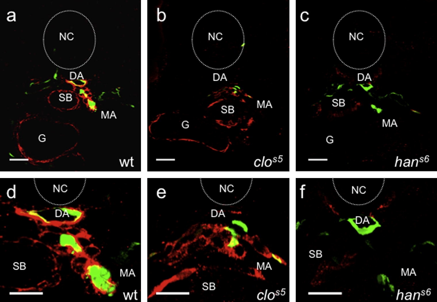
Fig. 6 Vascular mural cells derive from the lateral plate mesoderm but not from the blood or endothelial lineages. Confocal images of transverse sections, at different magnifications, of 80 hpf Tg(flk1:GFP)s843 wild-type (a and d) or cloche (clos5) (b and e) or hands-off (hans6) (c and f) mutant larvae analyzed for Transgelin expression (red). While wild-type (wt) and clos5 mutant larvae show Transgelin-positive cells around both the dorsal aorta and visceral organs, hans6 mutant larvae completely lack both vascular and visceral MCs. Sections are at the level of the1st-2nd somite. Scale bars, 20 μm. NT, neural tube; DA, dorsal aorta; SB, swim bladder; G, gut; MA, mesenteric artery.
Reprinted from Mechanisms of Development, 126(8-9), Santoro, M.M., Pesce, G., and Stainier, D.Y., Characterization of vascular mural cells during zebrafish development, 638-649, Copyright (2009) with permission from Elsevier. Full text @ Mech. Dev.