
Image
|
Figure Caption
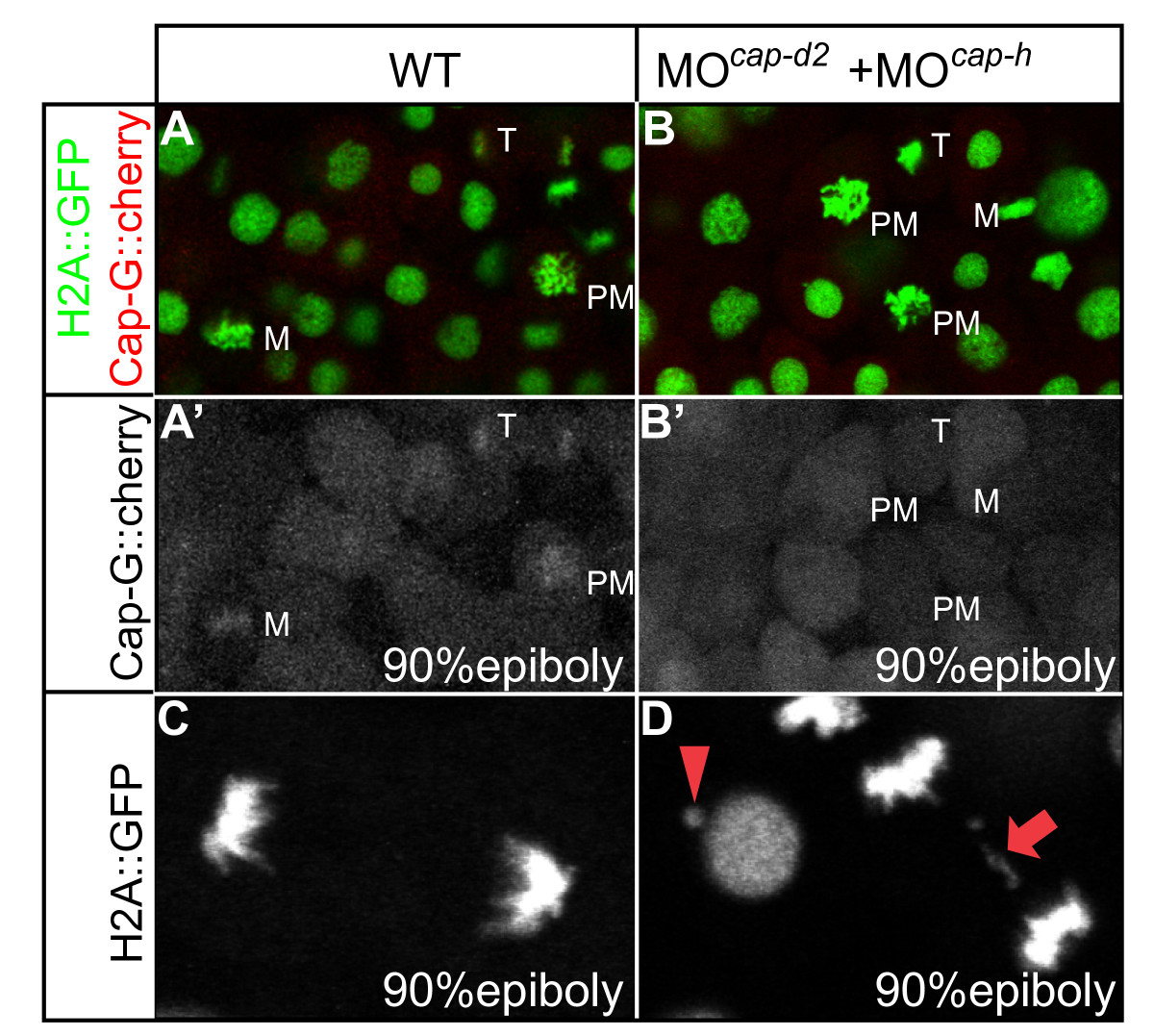
Fig. 7 CAP-G-mcherry is mislocalized during gastrula stage mitoses upon loss of its interaction partners CAP-H and CAP-D2 and localizes to the cytoplasm. (A, B) The different mitotic stages are recognizable by transgenic H2A::GFP expression. M, metaphase; PM, prometaphase; T, telophase. (C, D) Occurrence of chromatid bridges in MOcap-h+cap-d2 injected embryos. Red arrow indicates a chromatid bridge in between two anaphase nuclei. Red arrowhead marks aberrant genetic material associated with a decondensed nucleus.
Acknowledgments
This image is the copyrighted work of the attributed author or publisher, and
ZFIN has permission only to display this image to its users.
Additional permissions should be obtained from the applicable author or publisher of the image.
Full text @ BMC Dev. Biol.