
Image
|
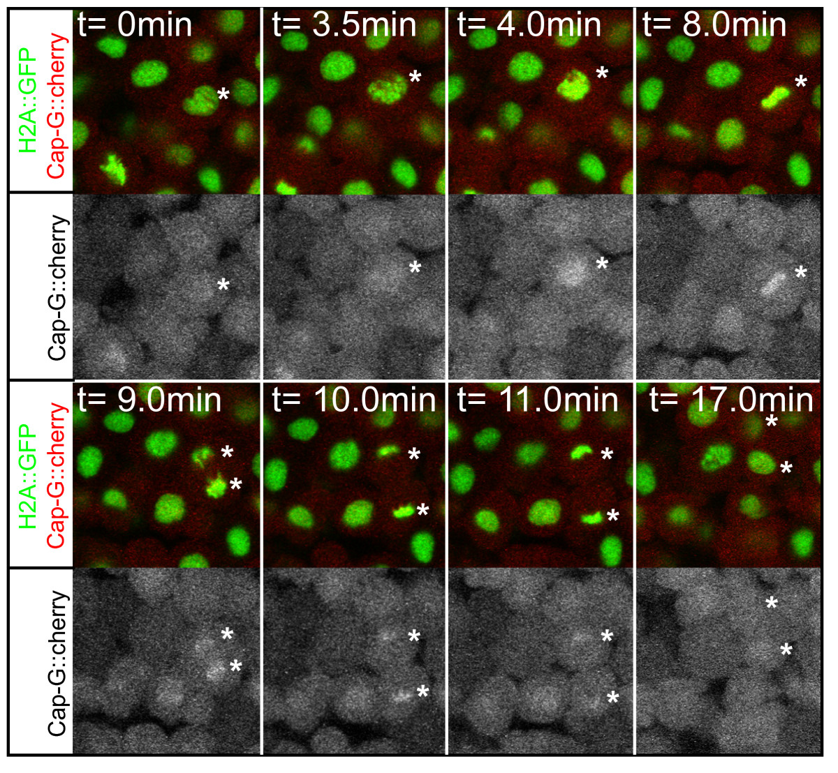
Figure Caption
Fig. 6 Chromatid association of CAP-G during mitosis. Selected images from a timelapse recording of a CAP-G-mcherry fusion protein in a gastrula stage transgenic embryo. The fusion protein associates with chromatids after the breakdown of the nuclear envelope at the beginning of prometaphase (t = 4.0 min) where it remains until decondensation of chromosomes during telophase (t = 17.0 min). Asterisks indicate positions of segregating chromatids. The different mitotic stages are recognizable by transgenic H2A::GFP expression. M, metaphase; PM, prometaphase; T, telophase.
Acknowledgments
This image is the copyrighted work of the attributed author or publisher, and
ZFIN has permission only to display this image to its users.
Additional permissions should be obtained from the applicable author or publisher of the image.
Full text @ BMC Dev. Biol.