
|
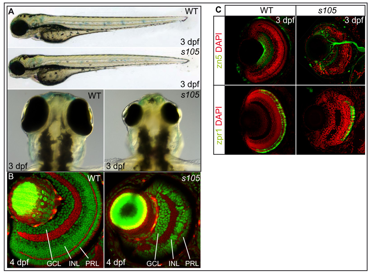
Fig. 1 s105 mutant embryos have a strong reduction in retinal cell number. (A) Wild-type (WT) and s105 mutant embryo at 3 dpf. The s105 mutant is characterized by small eyes. (B) Transverse vibratome sections through embryonic retinae counterstained with phalloidin to visualize plexiform layers (red) and propidium iodide to mark nuclei (green). In s105 mutants, the retina is smaller but some stratification of the retinal layers is present. (C) Transverse vibratome sections through embryonic retinae counterstained with DAPI to visualize nuclei (red), with the zn5 antibody to detect ganglion cells and the optic nerve (green), or with the zpr1 antibody to detect red/green double cones (green). GCL, ganglion cell layer; INL, inner nuclear layer; PRL, photoreceptor cell layer.