
Fig. 5
- ID
- ZDB-IMAGE-090817-46
- Publication
- Sun et al., 2009 - Cardiac hypertrophy involves both myocyte hypertrophy and hyperplasia in anemic zebrafish
- All Figures
- Figures for Sun et al., 2009
|
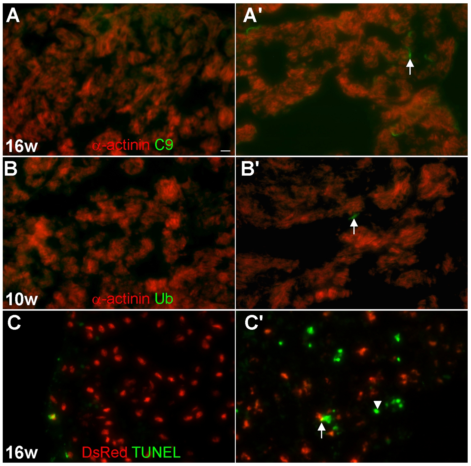
Fig. 5 Oncosis is the main form of cardiomyocyte death in tr265/tr265.
(A–A′) Increased number of cardiomyocytes undergoing oncosis in 10-μm ventricle sections of (A′) tr265/tr265 compared to (A) the sibling detected by a C9 antibody (green) and α-actinin antibody (red; cardiomyocytes); arrow points to a C9-positive cardiomyocyte; bar = 10 μm. (B,B′) Few, but more, autophagic cardiomyocytes seen in (B′) tr265/tr265 vs. (B) the sibling with a ubiquitin antibody (green) and α-actinin antibody (red; cardiomyocytes); arrow points to an ubiquitin-positive cardiomyocyte. (C,C′) Increased number of apoptotic non-cardiomyocytes (green overlay; arrowhead) in (C′) tr265/tr265, compared to (C) the sibling detected with TUNEL (green); cardiomyocytes (nuclear DsRed); arrowheads point to the rare apoptosing cardiomyocytes (yellow overlay).