
|
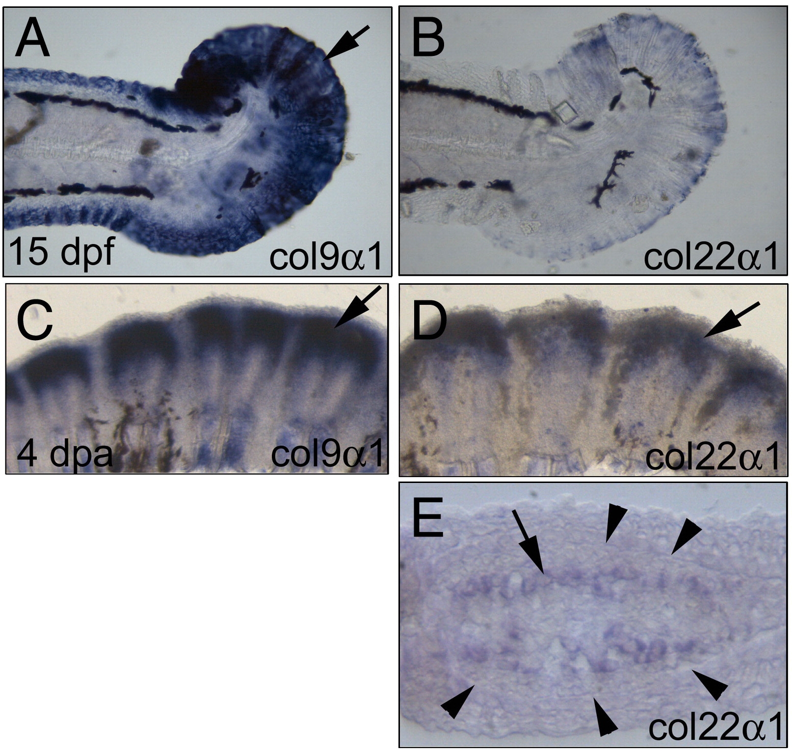
Fig. S2 Expression patterns of col9α1 and col22α1 in the fins. (A) col9α1 was highly expressed in the 15-day post-fertilization (dpf) fin (arrow). (B) col22α1 was also weakly expressed in the 15-dpf fin. (C) In a 4-day post-amputation (dpa) regenerating fin, col9α1 was expressed in large amounts in the regenerating fin (arrow). (D) The 4-dpa regenerating fin also expressed high levels of col22α1 but in a relatively smaller domain (arrow). (E) Cross-section of (D) revealing that col22α1 was expressed in osteoblasts located inside the fin ray. The number of cells expressing col22α1 (arrows) was apparently smaller than that expressing col9α1 (compare with Fig. 3H). Arrowheads denote the thick basement membrane (seen as a line at this magnification) where the future fin ray will form in the regenerating fin.
Reprinted from Developmental Biology, 332(2), Huang, C.C., Wang, T.C., Lin, B.H., Wang, Y.W., Johnson, S.L., and Yu, J., Collagen IX is Required for the Integrity of Collagen II Fibrils and the Regulation of Vascular Plexus Formation in Zebrafish Caudal Fins, 360-370, Copyright (2009) with permission from Elsevier. Full text @ Dev. Biol.