
|
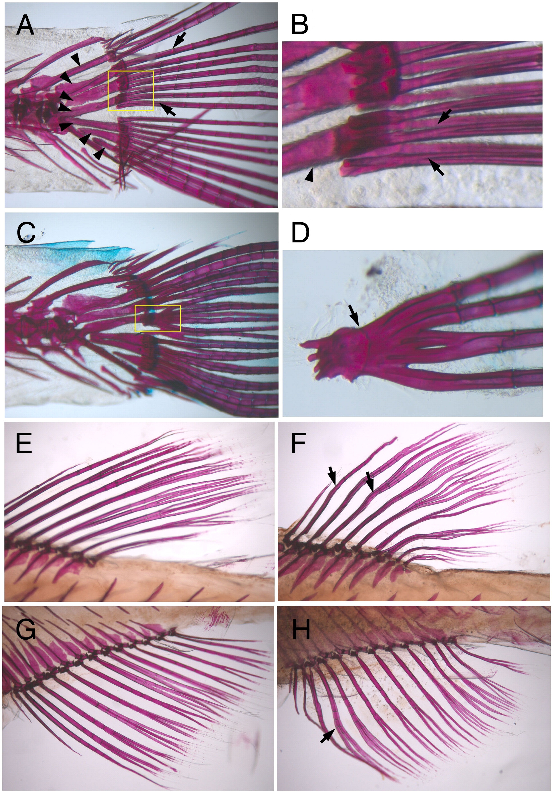
Fig. S1 Wavy and fused lepidotrichia in prp mutants. Alizarin red staining of ossified bones (red) in zebrafish. Regions of the fin base are shown. (A, B) Wild-type (WT) adult zebrafish showing that lepidotrichia (arrows) were connected to the body through radial plates (arrowheads). (B) Close view of the lepidotrichia bases (boxed region of A) showing that the lepidotrichia were closely yet separately positioned (arrows). Arrowhead, radial plate. (C, D) The lepidotrichia in the prp mutant were often fused at the base (arrows). Although they managed to grow out as separate fin rays, they might be fused at more-distal points later in development (Fig. 2P). Alizarin red staining of WT dorsal (E) and anal (G) fins and prp dorsal (F) and anal (H) fins showing that the lepidotrichia of prp fins were wavy (arrows).
Reprinted from Developmental Biology, 332(2), Huang, C.C., Wang, T.C., Lin, B.H., Wang, Y.W., Johnson, S.L., and Yu, J., Collagen IX is Required for the Integrity of Collagen II Fibrils and the Regulation of Vascular Plexus Formation in Zebrafish Caudal Fins, 360-370, Copyright (2009) with permission from Elsevier. Full text @ Dev. Biol.