
|
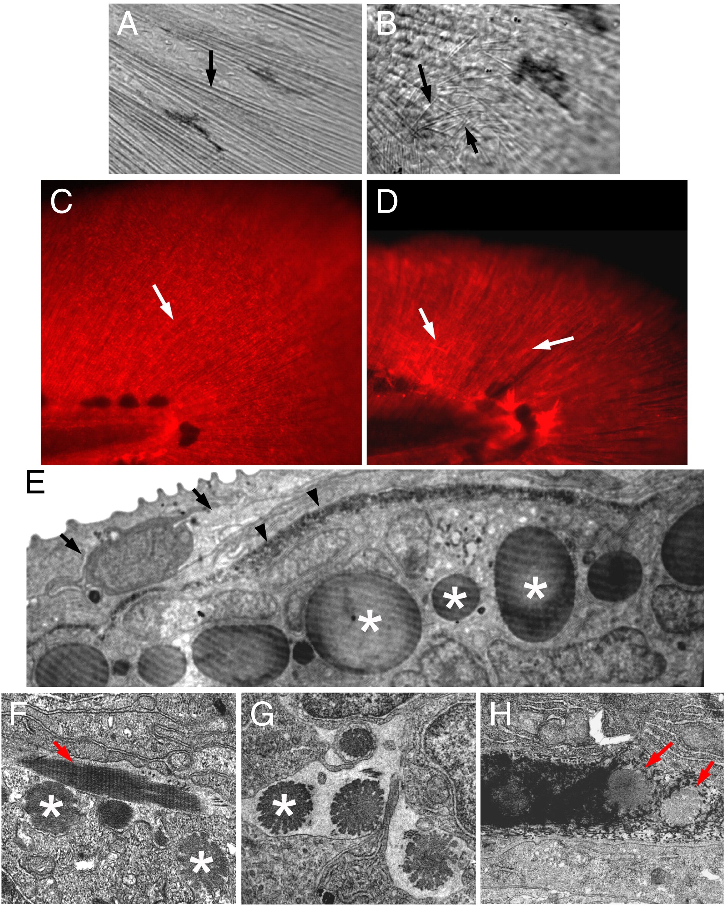
Fig. 6 Actinotrichia defects in prp mutants. Bright light examination of (A) wild-type (WT) and (B) prp finfolds revealed short and mis-orientated actinotrichia in prp (arrows). (C, D) Immunostaining of zebrafish finfolds with a monoclonal antibody against human collagen II. (C) In the WT finfold, this antibody stained the actinotrichia (arrows). (D) In prp, however, the actinotrichia appeared less dense and were often crisscrossed (arrows). (E) Analyses with transmission electronic microscopy (TEM) showed that in the WT fin, actinotrichia (asterisks) were bundles of collagen matrices located beneath the epidermis (arrow) and lepidotrichia (arrowheads). (F–H) The actinotrichia of the prp mutant appeared broken and much smaller (asterisks), sometimes perpendicular (arrow in panel F), and misassembled within the lepidotrichia (arrow in panel H).
Reprinted from Developmental Biology, 332(2), Huang, C.C., Wang, T.C., Lin, B.H., Wang, Y.W., Johnson, S.L., and Yu, J., Collagen IX is Required for the Integrity of Collagen II Fibrils and the Regulation of Vascular Plexus Formation in Zebrafish Caudal Fins, 360-370, Copyright (2009) with permission from Elsevier. Full text @ Dev. Biol.