
Image
|
Figure Caption
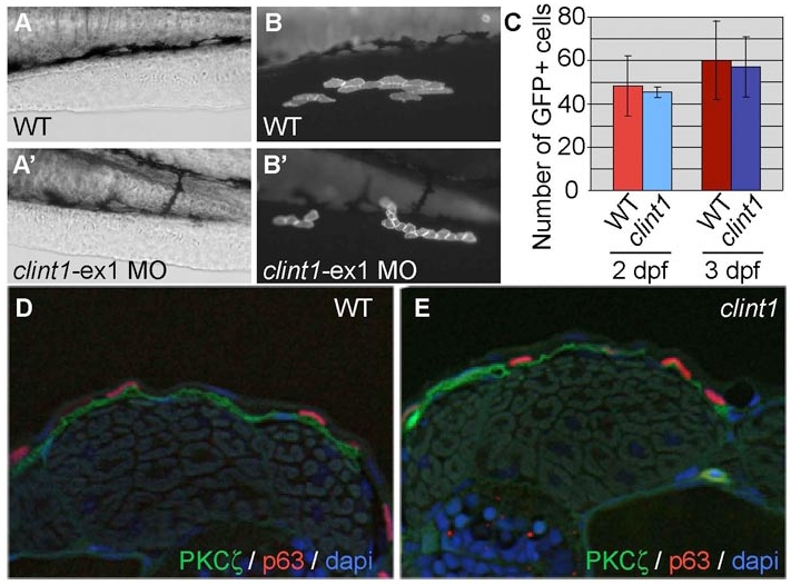
Fig. S6 Normal proliferation of clint1 morphant cells in a wild-type environment and unaffected Pkcζ localization in clint1 mutants. (A-B′) Cells from uninjected (A,B) or clint1-ex1 MO-injected (A′,B′) Tg(β-actin:hras-eGFP) embryos were transplanted to the ventral ectoderm of non-transgenic wild-type embryos. (C) Quantification of GFP-positive cells in unlabeled recipients at 2 and 3 dpf. (D,E) Transverse sections of wild-type (D) and clint1 mutant (E) embryos immunolabeled for Pkcζ (green) and p63 (red) and with DAPI (blue) at 48 hpf.
Acknowledgments
This image is the copyrighted work of the attributed author or publisher, and
ZFIN has permission only to display this image to its users.
Additional permissions should be obtained from the applicable author or publisher of the image.
Full text @ Development