
Image
|
Figure Caption
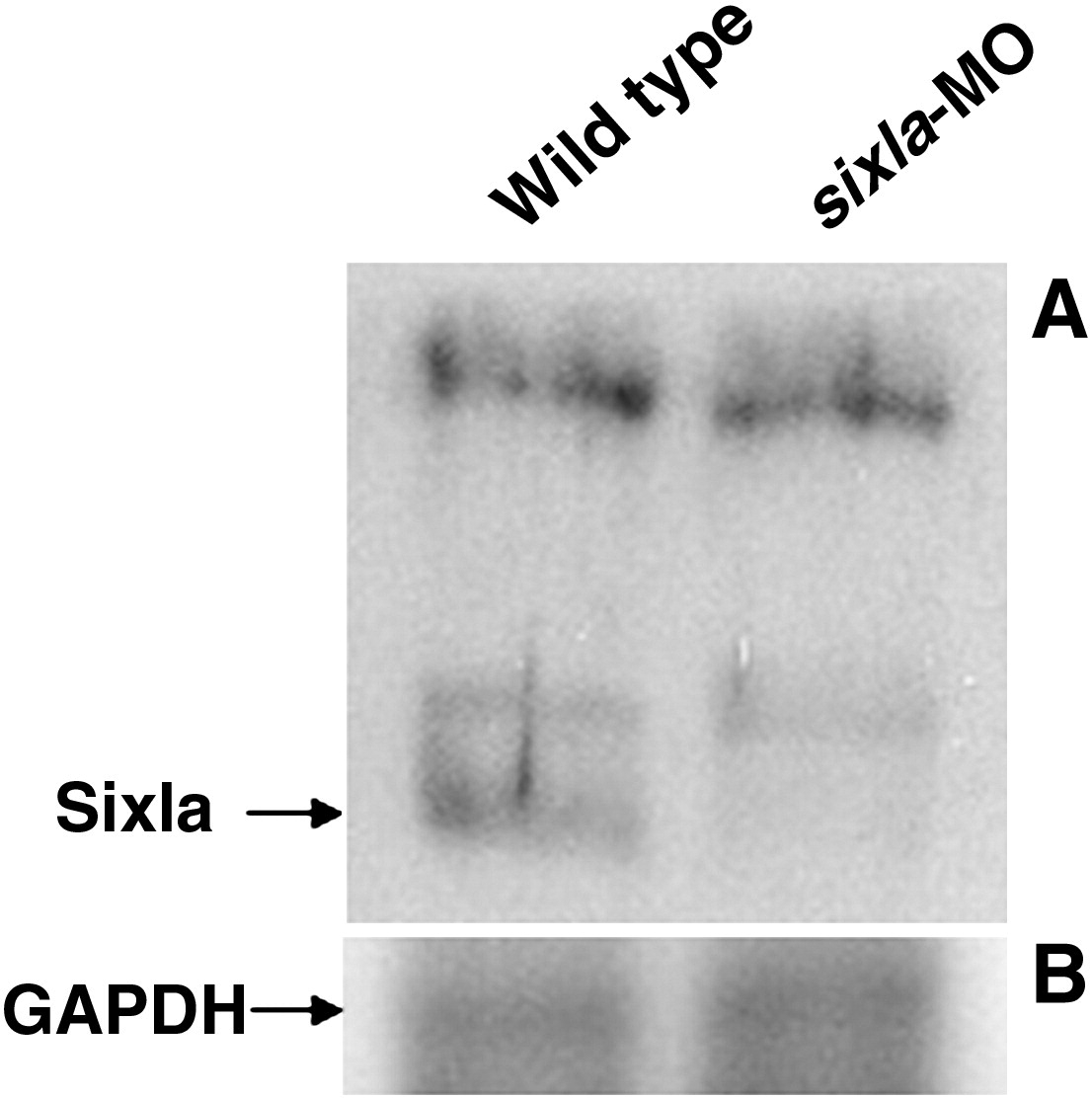
Fig. S2 Western blot analysis to detect the decrease of Six1a protein level in six1a morphants. Total protein lysates were extracted separately from the wild-type embryos (left lane) and from the six1a-MO-injected embryos (right lane) at 48 hpf. After the total proteins were separated on a 12% SDS-PAGE, polyclonal antibodies of anti-Six1a (panel A) and anti-Glyceraldehyde 3-phosphate dehydrogenase (GAPDH; panel B) were carried out for Western blot analysis at 1:1000 dilution. The intensity of positive band for antibody against GAPDH served as a protein-loading control.
Acknowledgments
This image is the copyrighted work of the attributed author or publisher, and
ZFIN has permission only to display this image to its users.
Additional permissions should be obtained from the applicable author or publisher of the image.
Reprinted from Developmental Biology, 331(2), Lin, C.Y., Chen, W.T., Lee, H.C., Yang, P.H., Yang, H.J., and Tsai, H.J., The transcription factor six1a plays an essential role in the craniofacial myogenesis of zebrafish, 152-166, Copyright (2009) with permission from Elsevier. Full text @ Dev. Biol.