
|
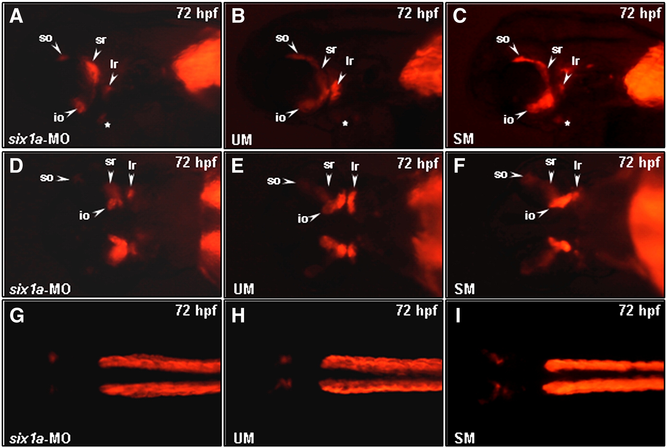
Fig. S1 Comparison of the muscle defects induced by injection of six1a-MO, UM-MO and SM-MO into embryos. Embryos derived from the transgenic line Tg (α-actin:RFP) (A–I) were injected with either 8 ng of six1a-MO (A, D, G), 8 ng of UM-MO (B, E, H) or 8 ng of SM-MO (C, F, I) to specifically inhibit the translation of six1a mRNA. RFP signal was detected only in the so, io, sr, lr, and remnant dorsal branchial arch muscle (white star) primordia in the six1a-, UM-and SM-MO-injected embryos at 72 hpf (A–F), whereas RFP was not detected in the trunk migratory muscle primordia in the six1a-, UM-and SM-MO-injected embryos at 72 hpf (G–I). Lateral view: A–C; ventral view: D–E; dorsal view: G–I. io, inferior oblique; ir, inferior rectus; lr, lateral rectus; so, superior oblique and sr, superior rectus.
Reprinted from Developmental Biology, 331(2), Lin, C.Y., Chen, W.T., Lee, H.C., Yang, P.H., Yang, H.J., and Tsai, H.J., The transcription factor six1a plays an essential role in the craniofacial myogenesis of zebrafish, 152-166, Copyright (2009) with permission from Elsevier. Full text @ Dev. Biol.