
|
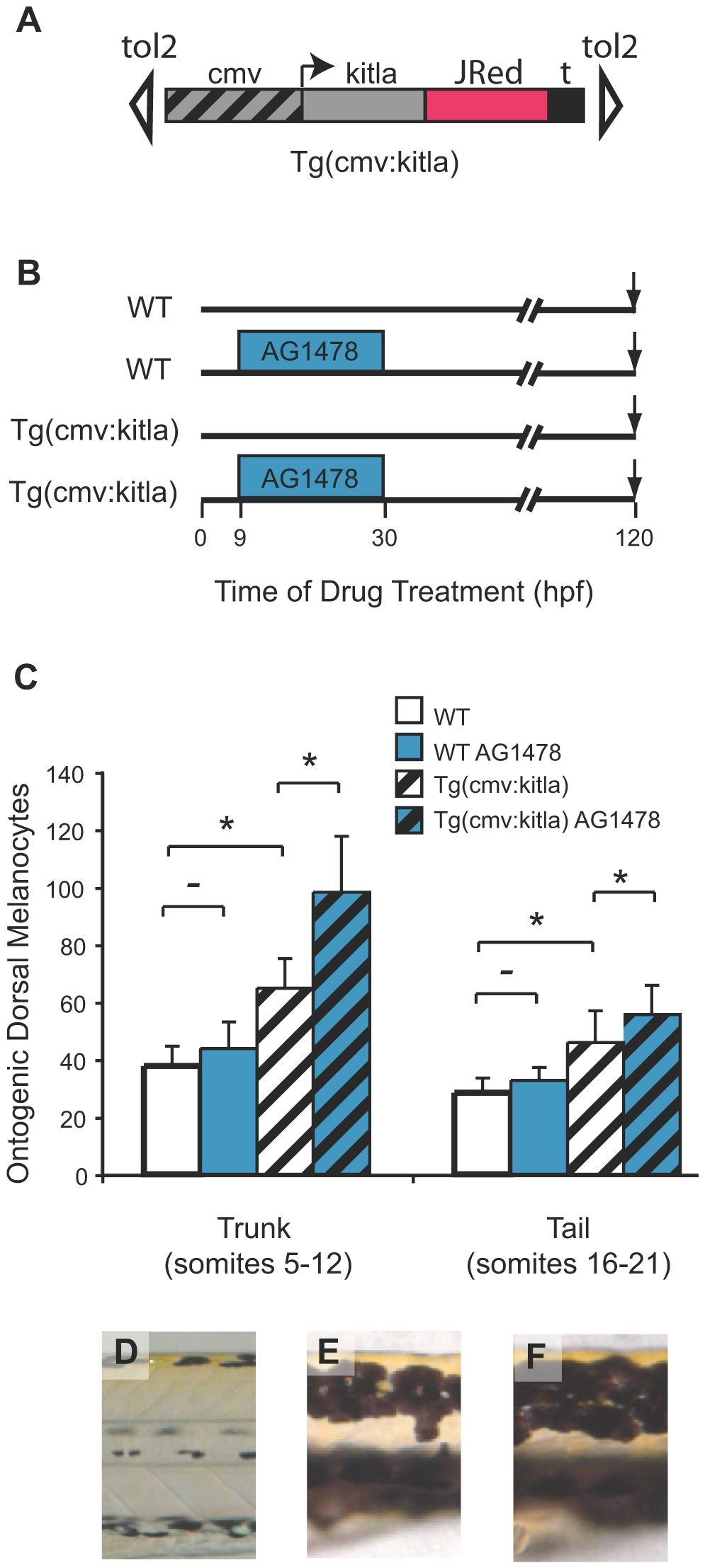
Fig. 7 kitla melanocyte proliferation is enhanced by AG1478.
(A) Cartoon of the PT2hsp70:kitla construct in transgenic animals. The constitutive cmv promoter drives expression of kitla. (B) Schematic of drug treatment timeline with early treatment of AG1478. (C) Quantitation of average ontogenetic dorsal melanocytes for each treatment in (A) in the trunk (somites 5–12) and in the tail (somites 16–21). Error bars represent standard deviation, * P<0.05, - P>0.05 (Student t-test, N = 10). As reported previously (see Figure 1) WT larvae show no effect in ontogenetic melanocyte number with AG1478 treatment compared to untreated WT. PT2hsp70:kitla animals have significantly more melanocytes in both the trunk and the tail. Treating PT2hsp70:kitla animals with AG1478 early, from 9–30 hpf, produces a significantly greater number of melanocytes in the trunk. In contrast, the tail region of AG1478-treated PT2hsp70:kitla larvae shows a significant, but much smaller increase than the trunk, in the number of excess melanocytes. (D) Trunk dorsal stripes of WT larvae are typically two melanocytes wide. (E) PT2hsp70:kitla, however, have trunk dorsal stripes that are about 4 cells wide. (F) AG1478-treated PT2hsp70:kitla have trunk dorsal stripes ∼6 melanocytes wide.