
|
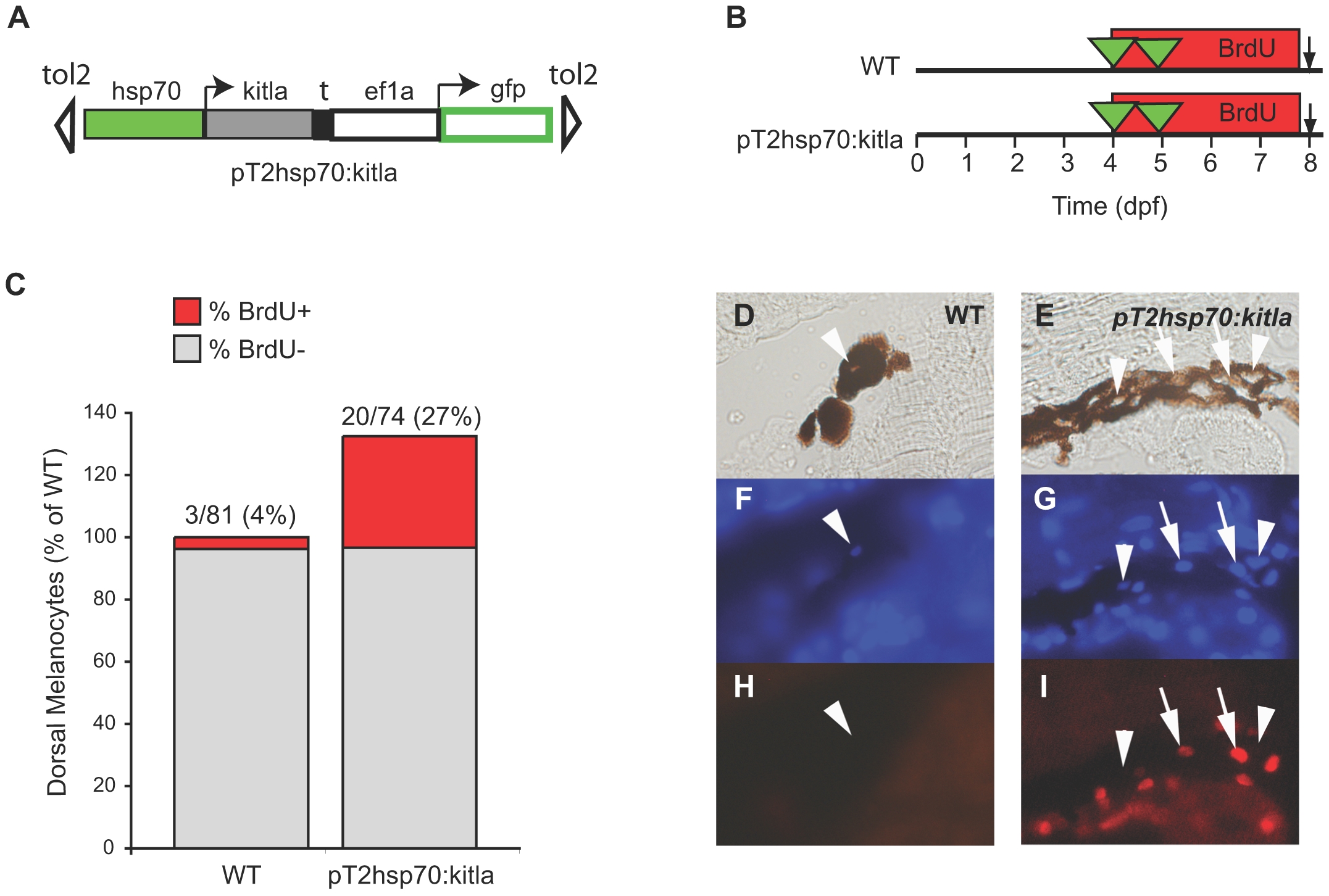
Fig. 5 Overexpression of kitla after 4 dpf results in proliferation of melanocytes.
(A) Cartoon of the pT2hsp70:kitla expression construct used in heatshock experiments. The heatshock promoter, hsp70 drives kitla expression, allowing for expression of kitla after heatshocking the injected embryos at 37°C. (B) Cartoon of experimental protocol. Following injection of pT2hsp70:kitla, larvae were heatshocked for 1 hour at 4 and 5 dpf in the presence of BrdU (from 4 to 8 dpf). (C) Quantitation of dorsal melanocytes represented as a percentage of wild type, and the percentage of BrdU labeled melanocytes is represented in red for each treatment. Larvae injected with pT2hsp70:kitla and heatshocked develop approximately 30% more melanocytes than uninjected larva. These larvae also show an increased percentage of BrdU labeled melanocytes (∼27%) that is comparable to the number of excess melanocytes. Representative examples of melanocytes in WT (D, F, H) and pT2hsp70:kitla (E, G, I), showing melanocyte nuclei stained with DAPI for WT (F) and pT2hsp70:kitla (G), and BrdU staining for WT (H) and pT2hsp70:kitla (I). Melanocytes considered having BrdU+ nuclei are labeled with arrows and BrdU- nuclei are labeled with arrowheads.