
|
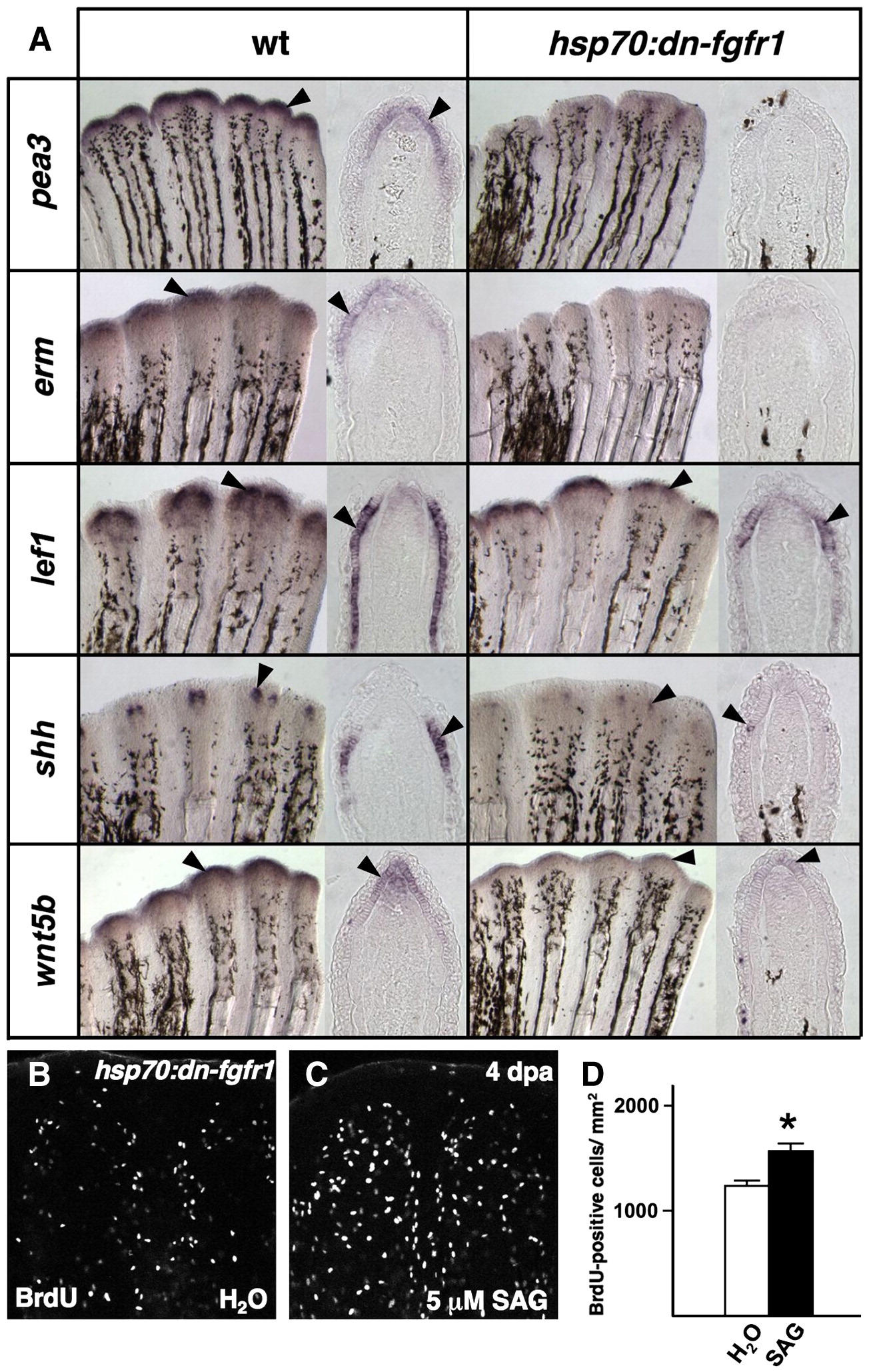
Fig. 5 Transgenic Fgfr blockade depletes both positive and negative epidermal regulators. (A) Whole-mount ISH for epidermal markers, and representative sections, 5 h after a single heat-shock in 4 dpa wildtype and hsp70:dn-fgfr1 regenerates. The expression of Fgf target genes pea3 and erm is markedly reduced during Fgfr inhibition, as are other epidermal markers lef1, shh, and wnt5b (arrowheads). (B, C) Images of BrdU incorporation in 4 dpa fin regenerates of heat-shocked hsp70:dn-fgfr1 animals treated with SAG. SAG augmented blastemal proliferation in hsp70:dn-fgfr1 animals. (D) Quantification of BrdU-positive cells in SAG- and vehicle-treated hsp70:dn-fgfr1 animals (n = 12; mean ± SEM; Student′s t-test, *P < 0.005).
Reprinted from Developmental Biology, 331(2), Lee, Y., Hami, D., De Val, S., Kagermeier-Schenk, B., Wills, A.A., Black, B.L., Weidinger, G., and Poss, K.D., Maintenance of blastemal proliferation by functionally diverse epidermis in regenerating zebrafish fins, 270-280, Copyright (2009) with permission from Elsevier. Full text @ Dev. Biol.