
|
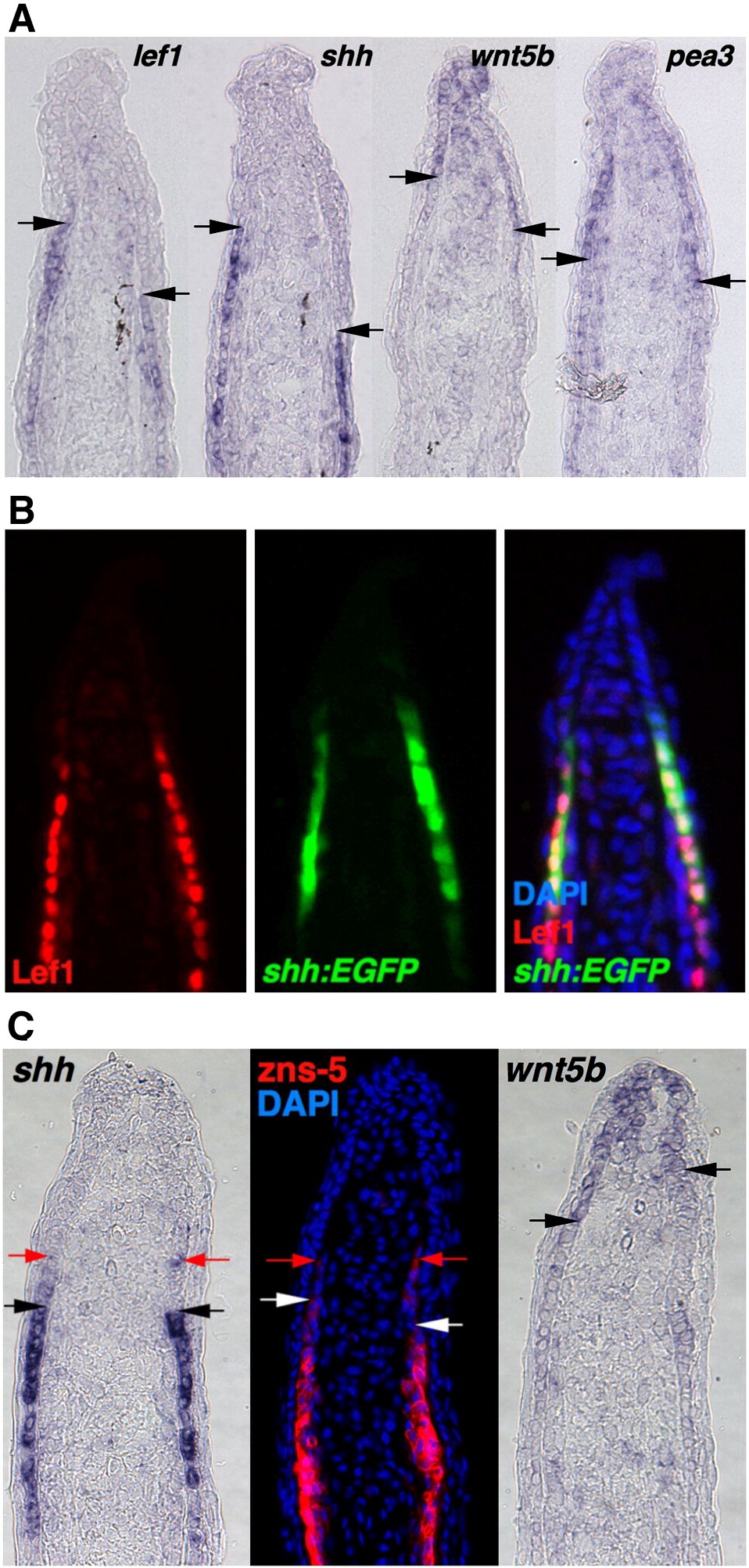
Fig. 2 The regenerating zebrafish fin has two epidermal cellular subtypes. (A) ISH for lef1, shh, wnt5b and pea3 in serial sections from a single 3 dpa (33 °C) fin regenerate. Arrows indicate the distal end of the lef1, shh or the proximal end of the wnt5b, pea3 signals. Note that section ISH (A) gives slightly different domain representation than whole-mount ISH as in Fig. 1A, based on differences in probe penetration and signal development (Smith et al., 2008). (B) Antibody staining for Lef1 in a section from 3 dpa (33 °C) shh:EGFP fin regenerates. The distal limit of the Lef1 staining (red) is aligned with the distal limit of Shh expression (green). (C) ISH for shh, wnt5b and antibody staining for zns-5 to mark scleroblasts, using serial sections from single 3 dpa (33 °C) fin regenerate. Black and white arrows indicate the distal end of the shh and zns-5 signal, or the proximal end of the wnt5b signal. Red arrows indicate distal limits of weak signals, still similar between shh and zns-5, and non-overlapping with wnt5b.
Reprinted from Developmental Biology, 331(2), Lee, Y., Hami, D., De Val, S., Kagermeier-Schenk, B., Wills, A.A., Black, B.L., Weidinger, G., and Poss, K.D., Maintenance of blastemal proliferation by functionally diverse epidermis in regenerating zebrafish fins, 270-280, Copyright (2009) with permission from Elsevier. Full text @ Dev. Biol.