
|
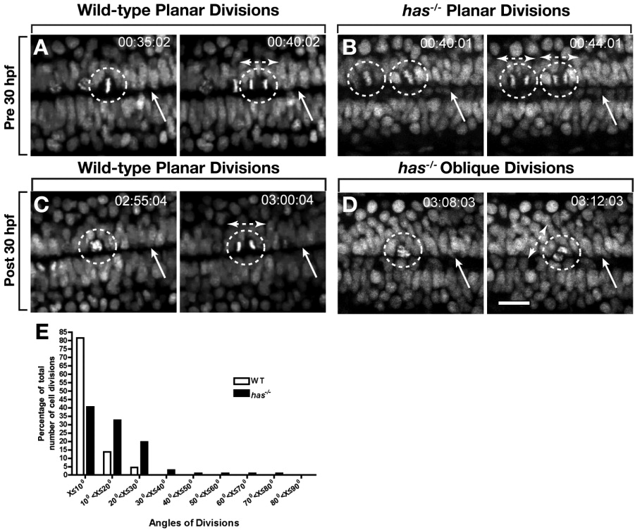
Fig. 2 Protein kinase C, iota (PrkCi) maintains planar divisions of cells that divide along the central canal. A-D: Frames captured from time-lapse movies, from a dorsal view, of wild-type and has-/- embryos carrying the Tg(h2afv:egfp) transgene. Numbers in upper right corners indicate time elapsed from beginning of imaging at 27 hours postfertilization (hpf). Dashed circles outline dividing cells, arrows point to the central canal and bi-directional arrows indicate orientation of the mitotic spindle and angle of division. A,B: In both wild-type and has-/- embryos, divisions that occur before 30 hpf are planar. The central canal is less distinct in has-/- embryos than in wild-type. C,D: Divisions after 30 hpf remain planar in wild-type embryos (C) but become oblique in has-/- embryos (D). The central canal is indistinct and appears to have been replaced by cells. E,F: Quantification of angles of division in wild-type and has-/- embryos (E). In wild-type most division planes are within 15° of the plane of the epithelium, indicated by the central canal. has-/- embryos have numerous divisions greater than 15°. Scale bar = 24 μM.