
|
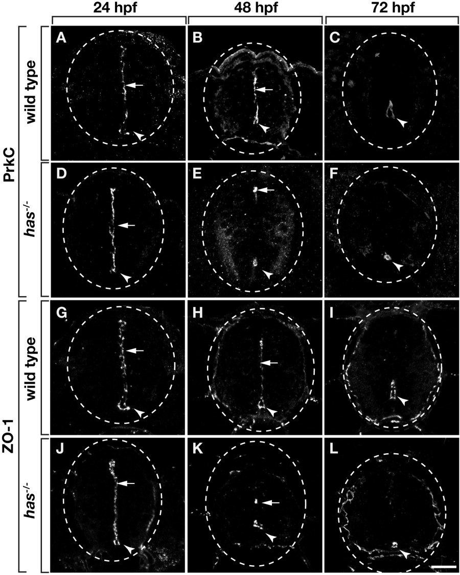
Fig. 1 Zebrafish spinal cord cells have apical polarity, which requires protein kinase C, iota (PrkCi) function. All panels show transverse sections through trunk spinal cord, dorsal up. Dashed circle marks the perimeter of the spinal cord. Arrowheads and arrows indicate central canal and medial septum, respectively. A-F: Sections labeled with anti-PrkCi/z antibody. A,B: PrkC is localized to the medial septum and central canal of wild-type embryos at 24 and 48 hours postfertilization (hpf). C: At 72 hpf, PrkC is absent from the medial septum but remains around the central canal of wild-type larvae. D: At 24 hpf, PrkC localization is normal in has-/- embryos. E: However, by 48 hpf very little PrkC is evident at the medial septum and, although PrkC remains around the central canal, the central canal is reduced in size (E) or entirely absent (not shown). F: Sections of 72 hpf has-/- larvae similarly reveal PrkC localization around an abnormally small and discontinuous central canal. The apical protein ZO-1 has a similar localization pattern to PrkC in wild-type and has-/- embryos and larvae (G-L). Scale bar = 20 μM.