
|
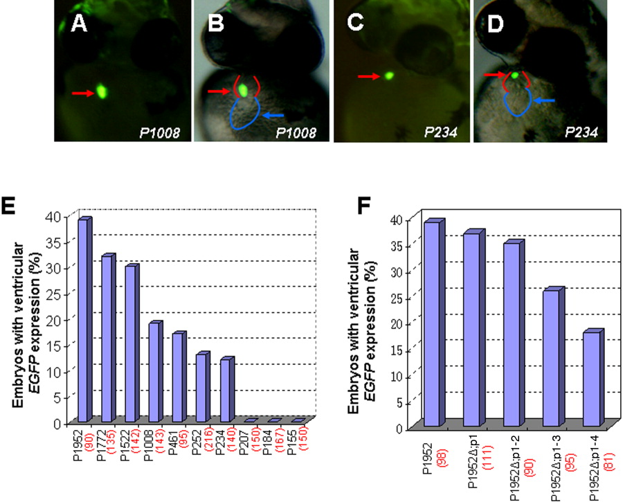
Fig. 3 Effects of vmhc promoter deletions on transgene EFFP expression in the ventricle. A-D: Embryos injected with a series of distal promoter deletion constructs at one- or two-cell stages were examined for transient EGFP expression at 48 hpf using fluorescent microscopy. Fluorescent optics showing ventricular EGFP expression in embryos injected with P1008 (A, B) and reduced EGFP expression in embryos with P234 injection (C, D). Reduced EGFP fluorescence typically correlates with a smaller vmhc promoter. Red and blue lines sketch the ventricle and atrium, respectively. E: Bar graph showing percentages of embryos that express EGFP in the ventricle in microinjection of each deletion construct. F: Bar graph depicting percentages of ventricular EGFP-expressing embryos injected with Prx2/S8-deletion constructs (P1952 Δp1, P1952 Δp1-2, P1952 Δp1-3, P1952Δ p1-4). The total number of embryos injected with each construct is shown in parentheses. Ventral views (A-D).