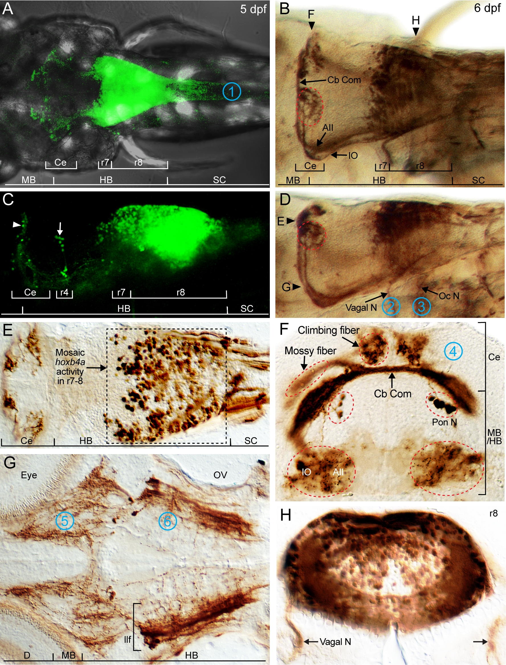
Fig. S1 Live imaging and immunohistochemically detected hoxb4a activity in the midbrain, cerebellum, hindbrain and spinal cord. Composite dorsal (A) and side (C) views of hoxb4a expression in a live 5 dpf transgenic zebrafish from 210 μm and 150 μm confocal stacks, respectively. (B, D) Dorsal (B) and side (D) views of hoxb4a-YFP using immunohistochemistry (anti-YFP) in a fixed 6 dpf fish. Horizontal (E, G) and coronal sections (F, H) with section planes indicated in (B, D). Target sites for retrograde labeling are marked by 1 (spinal cord), 2 (Xth nerve), 3 (pectoral fin), 4 (cerebellum), 5 (midbrain) and 6 (r4). Abbreviations: AII, Area II; Ce, cerebellum; D, diencephalon; HB, hindbrain; llf, lateral longitudinal fascicle; IO, inferior olive; MB, midbrain; mlf, medial longitudinal fascicle; Oc N, occipital nerve; OV, otic vesicle; Pon N, pontine nucleus; SC, spinal cord; Vagal N, vagal nerve. B, D and E–H are cropped high magnification illustrations of Figs. 5K, 5J, 5C, 5T, 5D and 4B, respectively, (from [25]).
Image
Figure Caption
Acknowledgments
This image is the copyrighted work of the attributed author or publisher, and
ZFIN has permission only to display this image to its users.
Additional permissions should be obtained from the applicable author or publisher of the image.
Full text @ PLoS One

