
|
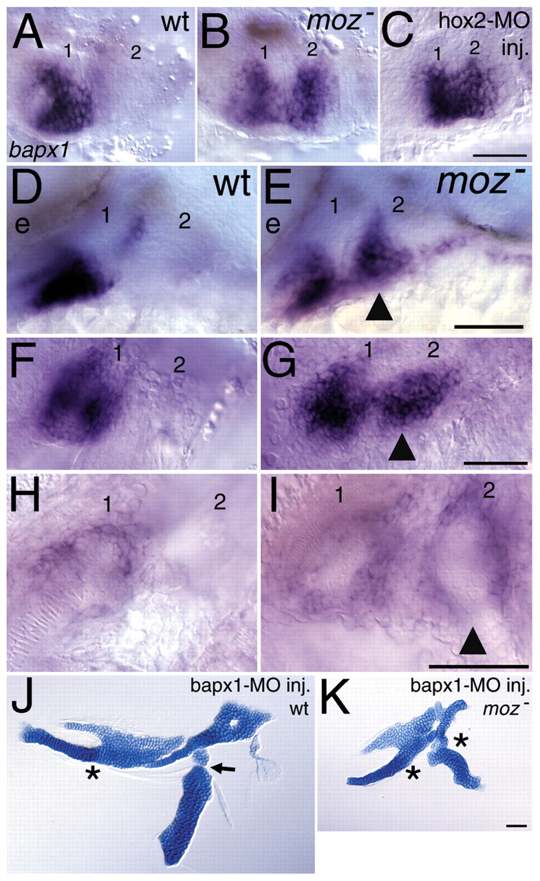
Fig. 9 Stable ectopic expression of bapx1 in the moz mutant second arch. Ventral (A-C,F-I) and lateral (D,E) views of bapx1 expression in whole-mount wild-type (A,D,F,H), moz mutant (B,E,G,I) and hoxa2b + hoxb2a-MO co-injected animals (C) at 33 hpf (A-E), 54 hpf (F,G) and 4 days (H,I). (A-E) bapx1 expression, which is normally restricted to a patch of first arch mesenchyme (A,D), is ectopically expressed (arrowhead in E) in the second arch of moz mutants (B,E) and hoxa2b+hoxb2a-MO co-injected animals (C). (F-I) Ectopic bapx1 expression (arrowheads) is maintained in second arch mesenchyme of moz mutants. (J,K) Reduction of bapx1 function in wild type (J) and moz mutant (K). Reducing bapx1 function in wild type causes specific loss of the jaw joint (asterisk in J), while reducing function of bapx1 in moz mutants causes loss of both first and second arch joints (asterisks in K). Reduction of bapx1 function in moz mutants also can rescue the ventral arch one and two fusions. The first two pharyngeal arches are numbered. e, eye. Scale bars: 50 μm.