
|
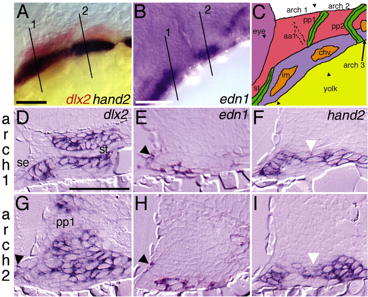
Fig. 1 edn1 pharyngeal arch expression is ventrally confined. Lateral views of (A) dlx2 and hand2 expression in red and blue, respectively, at 28 hpf and (B) edn1 expression at 36 hpf. (C) Schematic of zebrafish pharyngeal arch primordia from 28-36 hpf at a slightly dorsal-oblique lateral view. The first and second arch ventral myogenic arch cores (intermandibularis and constrictor hyoideus ventralis; shown in orange) (see Kimmel et al., 2001b) express edn1 (Miller et al., 2000). The third arch myogenic core also expresses edn1. Pharyngeal epithelia, the stomodeum and pharyngeal pouches, are colored green. The first and second arches are labeled over the arches, with the postmigratory cranial neural crest (CNC) colored in red (dlx2+dHAND-) or blue (dlx2+dHAND+). Approximate section planes for the first two arches are indicated and numbered (A,B) or marked with arrowheads (C). (D-I) Transverse sections through the first two arches of 32 hpf embryos stained for dlx2 (D,G), edn1 (E,H) or hand2 (F,I). hand2-expressing cells are a ventral subset of dlx2-expressing CNC cells, including cells just dorsal (white arrowheads) to the edn1-expressing ventral arch cores. Lateral surface ectoderm expresses both dlx2 and edn1 ventrally (arrowheads). chv, constrictor hyoideus ventralis; im, intermandibularis; pp1, pharyngeal pouch 1; se, surface ectoderm; st, stomodeum. Scale bars: 50 μm.