
Image
|
Figure Caption
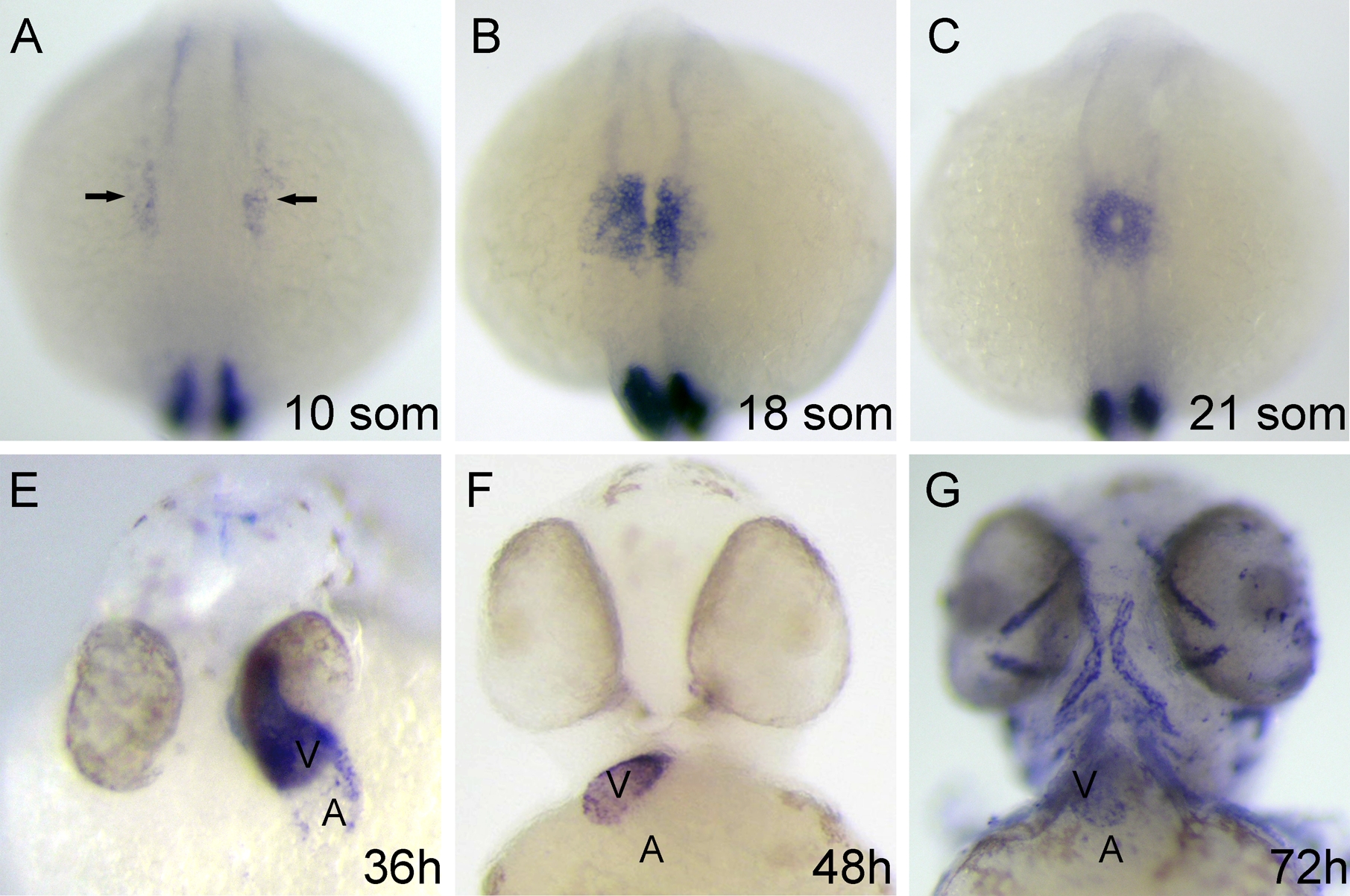
Fig. S1 vmhc gene expression profile. In situ hybridization results of embryos at various stages. The MyoD riboprobe was included in A-C to ensure proper staging. The vmhc transcript could be detected in the anterior lateral plate mesoderm as early as the 10-somite stage (A). Expression persists in the heart progenitor region, which migrates toward the midline (B), fuses to form a heart tube (C), and differentiates into two distinct chambers (D-F). vmhc expression can be detected in the extraocular muscles and pharyngeal muscles after 3 dpf (F).
Acknowledgments
This image is the copyrighted work of the attributed author or publisher, and
ZFIN has permission only to display this image to its users.
Additional permissions should be obtained from the applicable author or publisher of the image.
Full text @ Dev. Dyn.