
|
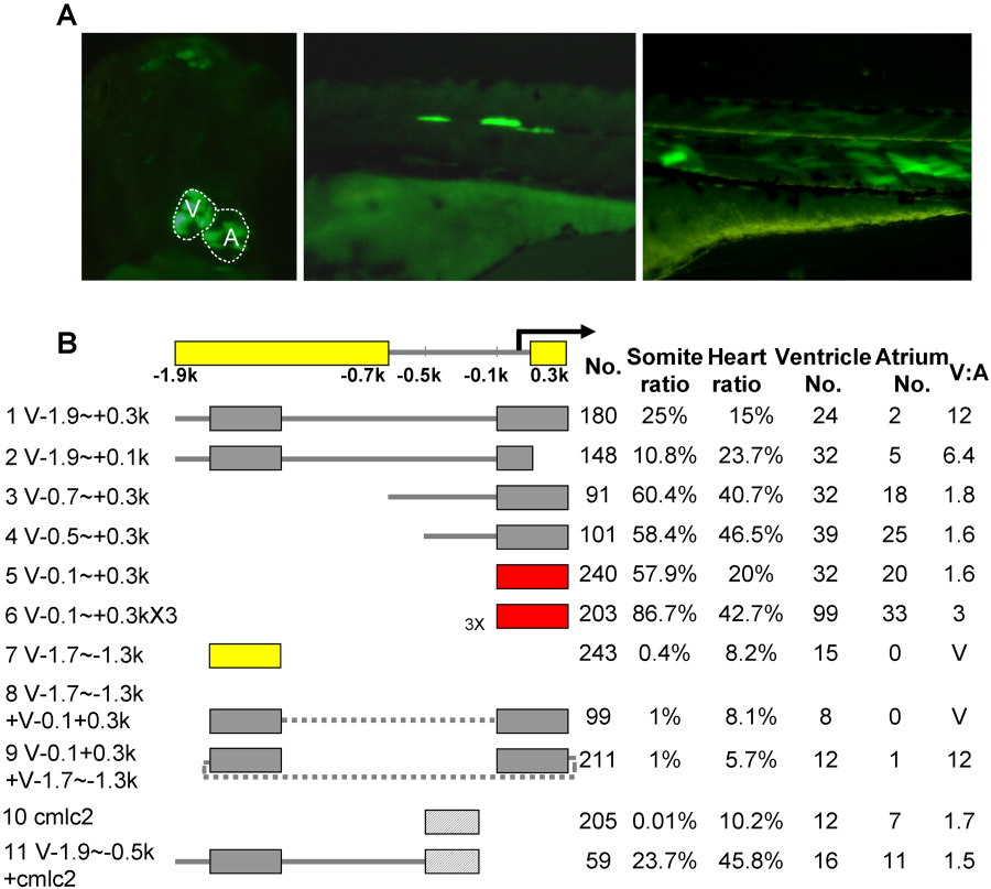
Fig. 3 Dissection of the vmhc promoter by transient assays using Tol2-based vectors. A: Representative pictures of 3-dpf embryos injected with Tol2 transposon constructs containing vmhc promoter sequences (left and right panel, V-0.5∼+0.3k; middle panel, V-1.9∼+0.3k). Shown in the left panel is a ventral view with anterior to the top; right and middle panels are lateral views with anterior to the left. Multiple GFP-positive cells could be detected in the heart (left) and/or skeletal muscle, including the eye muscle, muscle pioneer cells located in the body midline (middle), and myocytes in the somites (right). B: Schematic summary of results from transient assays using the Tol2 transposon system. A distal (V-1.9∼-0.7k) and a proximal (V+0.1∼+0.3k) element required for the chamber specificity were identified, consistent with results from transient co-injection assays in Figure 2. A shorter distal element (V-1.7∼-1.3k, line 7) is sufficient to drive ventricle-specific expression in this assay. In contrast to results from transient co-injection assays, the proximal element (V-0.1∼+0.3k, line 5) drives GFP expression in the whole heart without chamber specificity in Tol2-based assays. When the distal and proximal elements are linked in tandem (line 8) or in reverse (line 9), the constructs drive GFP expression in the ventricle. However, the distal element cannot alter the expression of the cardiac cmlc2 promoter to be chamber-specific (line 11), which by itself drives GFP expression in the whole heart (line 10). The minimal elements required for chamber specificity (yellow bars, top line), for chamber-restricted expression (yellow bars, line 7) and for cardiac expression (red bars, lines 5-6) are indicated. No., number of injected embryos that survived to 4 dpf; Somite ratio/Heart ratio, number of fish with tissue-restricted GFP-positive cells over total number of fish that survived to 4 dpf; Ventricle No. or Atrium No., number of fish with GFP-positive cells in the ventricle or atrium. A fish with GFP-positive cells in both chambers was counted in both categories. V:A, ratio of GFP-positive cells in the ventricle over that in the atrium.