
|
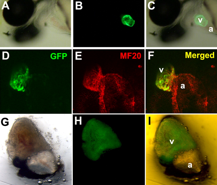
Fig. 3 Ventricle-specific expression of the green fluorescent protein (GFP) reporter gene using the 5′-flanking region of vmhc as a promoter. Transgenic zebrafish embryo at 48 hours postfertilization (hpf). A: Nomarski image. B: Ventricle-specific green fluorescent signal. C: Overlay. Confocal-microscopic (Zeiss) images of the 48-hpf transgenic embryo immunostained with myocardium-specific MF20 monoclonal antibody followed by the Alexa594 secondary antibody. D-F: Ventral heart view with green fluorescent signal at the ventricular region (D), red (E), overlay (F). G: Isolated heart from 2 months after fertilization using a Leica fluorescence stereomicroscope. H,I: Brightfield, green fluorescent signal in the ventricular region (H), overlay (I). Views of the embryos are lateral and anterior to the left. a, atrium; v, ventricle.