
|
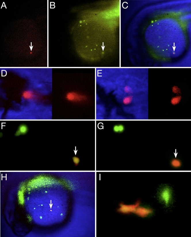
Fig. 4
Not All Blood Is Committed to a Unipotential Lineage at 26 hr
(A–E) Single circulating blood cells give rise to neutrophil and erythrocyte progeny. (A–C) A newly photoconverted blood cell (arrow), within a ventral gastrula-derived clone. (A) Red wavelength, (B) green wavelength (note the other blood cells), and (C) composite image. (D and E) The resulting red-labeled clone at 48 hr included (D) circulating erythrocytes and (E) two neutrophils attached to the lumen of a blood vessel.
(F–I) Single macrophage cells give rise to macrophage progeny. (F–H) High-magnification view of an individual macrophage (arrow) in the process of being photoconverted from (F) green to (G) red. Nearby, another green-labeled macrophage (upper left panel) is preparing to divide. (H) Low-magnification view of the entire Kaede clone showing the newly photoconverted macrophage (arrow) among other green-labeled macrophages and out-of-focus endoderm and endothelium. (I) The resulting red-labeled clone at 48 hr was solely two macrophages near a blood vessel. Another green-labeled macrophage is out of focus.
Reprinted from Developmental Cell, 16(5), Warga, R.M., Kane, D.A., and Ho, R.K., Fate mapping embryonic blood in zebrafish: multi- and unipotential lineages are segregated at gastrulation, 744-755, Copyright (2009) with permission from Elsevier. Full text @ Dev. Cell