
|
Fig. S4
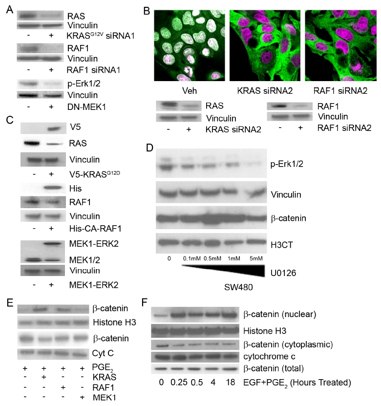
KRAS/RAF1 and COX-2/PGE2 Are Required to Maintain Nuclear Localization of β-Catenin in Human Cells
(A) SW-480 cells were transfected with control, KRASG12V- or RAF1-direceted siRNA or a dominant negative MEK1 construct. Whole cell protein was harvested and subjected to western blot analysis for total RAS and vinculin (top), total RAF and vinculin (middle) or pERK and vinculin (bottom). (B) SW-480 cells were transfected with KRAS or RAF1-directed siRNA constructs. Two days after transfection, the cells were fixed and stained for β-catenin (green) or ToPro3 (magenta) or whole cell lysates were harvested and subjected to western blot analysis for total RAS (top – left), total RAF1 (top – right) or vinculin (bottom). (C) SW-480 cells were transfected with KRASG12V-directed siRNA and transfected with constitutively active V5-KRAS, 6xHis-RAF1 or MEK1-ERK2 fusion. Whole cell lysates were harvested 48 hours after transfection and subjected to western blot analysis for V5 (top), total RAS (middle) and vinculin (bottom), 6xHis (top), total RAF1 (middle) and vinculin (bottom), or MEK1 (top) and vinculin (bottom). (D) SW-480 cells were treated with a dose curve (0.1μM, 0.5μM, 1μM or 5μM) of the MEK-specific inhibitor U0126. Following 6 hrs of treatment, whole cell protein and nuclear fractions were harvested and subjected to western blot analysis for whole cell pERK (top) and vinculin (2nd row) or nuclear β- catenin (3rd row) and histone H3 (bottom). (E) 293 cells transfected with VEH, constitutively active, KRAS, RAF1 or MEK1 and treated for 6 hrs with PGE2. Subcellular fractionation was performed and the fractions were subjected to western blot analysis for β-catenin (nuclear-top) or histone H3 or β-catenin (cytoplasmic-bottom) and cytochrome C. (F) 293 cells were treated with both PGE2 and EGF for 0, 0.25, 0.50, 4 or 18 hrs. Following treatment, subcellular fractionation was performed and the fractions were subjected to western blot analysis for β-catenin (nuclear-top) or histone H3 or β-catenin (cytoplasmic-bottom) and cytochrome C. Total β-catenin was also assayed from whole cell lysate. All images are representative of at least three individual experiments.
Reprinted from Cell, 137(4), Phelps, R.A., Chidester, S., Dehghanizadeh, S., Phelps, J., Sandoval, I.T., Rai, K., Broadbent, T., Sarkar, S., Burt, R.W., and Jones, D.A., A two-step model for colon adenoma initiation and progression caused by APC loss, 623-634, Copyright (2009) with permission from Elsevier. Full text @ Cell